RMF Logo

Article filters

Search Papers:

For searching before 2024 see Archive (2000-2023)
  • Vol. 2025, 43(4)
  • Scientific Article
  • Special Issue

Wheat leaf blight-associated fungi and their biological control

By Mariana Guadalupe Sánchez Alonso, Patricia Rivas Valencia*, Leticia Robles Yerena, Raúl Asael Rodríguez Villareal, María Florencia Rodríguez García, Raúl Rodríguez Guerra

*Corresponding Author.

Received: 18/November/2024 – Published: 26/June/2025
DOI: https://doi.org/10.18781/R.MEX.FIT.2024-26

  • PDF
  • HTML
  • XML

Abstract Background/Objective. The objective was to determine the inhibitory effect of actinobacterial morphotypes on mycelial growth of fungi associated with wheat leaf blight.

Materials and methods. A total of 136 samples of wheat leaves were processed from the highlands of Mexico. They were sectioned, washed and placed in a humid chamber; fungi were isolated on potato dextrose agar. A morphological identification was carried out and the incidence of diseases was recorded by federal entity. Actinobacteria were isolated from soil samples using the dilution technique and microcolonies were isolated on nutrient agar. Preliminary in vitro bioassays were established at 27 °C with 144 actinobacteria and a dual confrotation with morphotypes presented a percentage of inhibition of mycelial growth (PIMG) greater than 40% and to identify those with potential for the control of fungi associated with leaf spots. The experimental design was completely randomized and an analysis of variance (p ≤ 0.05) and mean test (Tukey p ≤ 0.05) were performed.

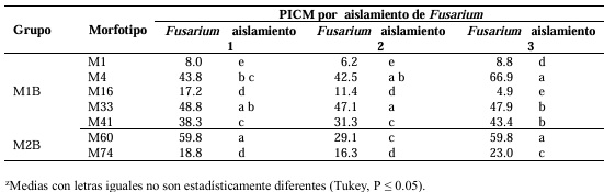
Results. At least three actinobacteria inhibited mycelial growth of Fusarium spp. in a range of 42.5 - 66.9 %. The inhibitory efficacy of strain M2B M60 was highest with Fusarium isolate 1, that of M1B33 with Fusarium isolate 2 and that of M1B M4 was most effective with Fusarium isolate 3.

Conclusion. The use of actinobacteria represents an alternative for integrated management in the control of fungi associated with foliar blight in wheat, as they present an inhibitory effect on mycelial growth.

Keywords: Actinobacteria, Fusarium, Specificity, Metabolites
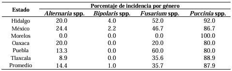
Table 1. Percentage of incidence of fungi associated with diseases in wheat crops sampled in the main producing states.
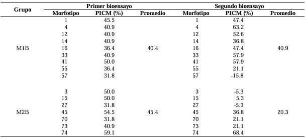
Table 2. Percentage inhibition of mycelial growth (PIMG) of preliminary bioassays of actinobacterial morphotypes against <em>Fusarium</em> isolate 1.
Table 2. Percentage inhibition of mycelial growth (PIMG) of preliminary bioassays of actinobacterial morphotypes against Fusarium isolate 1.
Table 3. Percentage inhibition of mycelial growth (PIMG) of three <em>Fusarium</em> isolates from wheat confronted with seven actinobacteria morphotypes.
Table 3. Percentage inhibition of mycelial growth (PIMG) of three Fusarium isolates from wheat confronted with seven actinobacteria morphotypes.
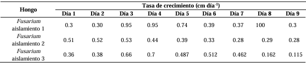
Table 4. Growth rate of <em>Fusarium</em> isolates from wheat leaves.
Table 4. Growth rate of Fusarium isolates from wheat leaves.
Figure 1. <em>Alternaria</em> spp. colonies. A) Colony on PDA culture medium. B) Colony on APZ culture medium. C) Early-stage mycelial growth on PDA culture medium. D) Colony on PDA medium showing one or two concentric rings observed under backlighting. E) Colony on PZA medium showing several concentric rings observed under backlighting. F-I) Conidia and conidial chains.
Figure 1. Alternaria spp. colonies. A) Colony on PDA culture medium. B) Colony on APZ culture medium. C) Early-stage mycelial growth on PDA culture medium. D) Colony on PDA medium showing one or two concentric rings observed under backlighting. E) Colony on PZA medium showing several concentric rings observed under backlighting. F-I) Conidia and conidial chains.
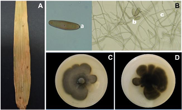
Figure 2. Symptoms and colonies of <em>Bipolaris</em> sp. A) Symptom of <em>Bipolaris</em> sp., of wheat leaf blight. B) Conidium with three slightly marked septa (a. basal scar), conidiophores (b), and mycelium (c). C) Colony on PDA culture medium. D) Reverse side of the Petri dish on PDA culture medium.
Figure 2. Symptoms and colonies of Bipolaris sp. A) Symptom of Bipolaris sp., of wheat leaf blight. B) Conidium with three slightly marked septa (a. basal scar), conidiophores (b), and mycelium (c). C) Colony on PDA culture medium. D) Reverse side of the Petri dish on PDA culture medium.
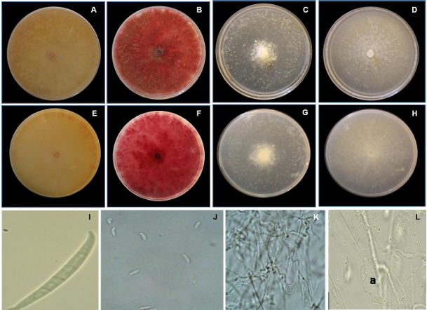
Figure 3. Characteristics of <em>Fusarium</em> spp. A and E) Front and reverse view of the colony on PDA culture medium of <em>Fusarium</em> isolate 1. B and F) Front and reverse view of the colony on SNA culture medium of <em>Fusarium</em> isolate 1. C and G) Front and reverse view of the colony on PDA culture medium of <em>Fusarium</em> isolate 2. D and H) Front and reverse view of the colony on SNA culture medium of <em>Fusarium</em> isolate 2. I) Macroconidium of <em>Fusarium</em> isolate 1. J) Microconidia of <em>Fusarium</em> isolate 2. K) Chlamydospores of <em>Fusarium</em> isolate 2. L) Microconidia and phialides (a) of isolate 2.
Figure 3. Characteristics of Fusarium spp. A and E) Front and reverse view of the colony on PDA culture medium of Fusarium isolate 1. B and F) Front and reverse view of the colony on SNA culture medium of Fusarium isolate 1. C and G) Front and reverse view of the colony on PDA culture medium of Fusarium isolate 2. D and H) Front and reverse view of the colony on SNA culture medium of Fusarium isolate 2. I) Macroconidium of Fusarium isolate 1. J) Microconidia of Fusarium isolate 2. K) Chlamydospores of Fusarium isolate 2. L) Microconidia and phialides (a) of isolate 2.
Figure 4. Rust pustules. A) Rust pustules on a wheat leaf. B) Urediniospore of <em>Puccinia</em> 1. C) Urediniospore of <em>Puccinia</em> 2. D) Teliospore of <em>Puccinia</em> 1. E) Teliospore of <em>Puccinia</em> 2.
Figure 4. Rust pustules. A) Rust pustules on a wheat leaf. B) Urediniospore of Puccinia 1. C) Urediniospore of Puccinia 2. D) Teliospore of Puccinia 1. E) Teliospore of Puccinia 2.
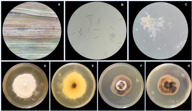
Figure 5. A) Chlorotic halos with pycnidia. B) Conidia of <em>Septoria</em> sp. C) Conidia of <em>Zymoseptoria</em> sp. observed under contrast phase. D) Front view of <em>Septoria</em> sp. colony on PDA. E) Reverse view of <em>Septoria</em> sp. colony on PDA. F) Front view of <em>Zymoseptoria</em> sp. colony on PDA. G) Reverse view of <em>Zymoseptoria</em> sp. colony.
Figure 5. A) Chlorotic halos with pycnidia. B) Conidia of Septoria sp. C) Conidia of Zymoseptoria sp. observed under contrast phase. D) Front view of Septoria sp. colony on PDA. E) Reverse view of Septoria sp. colony on PDA. F) Front view of Zymoseptoria sp. colony on PDA. G) Reverse view of Zymoseptoria sp. colony.
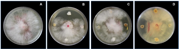
Figure 6. <em>Fusarium</em> isolate 1, nine days old. A) Control. B) and C) Interaction of the fungus with four actinobacteria, front view. D) Interaction of the fungus with four actinobacteria, back of the Petri dish.
Figure 6. Fusarium isolate 1, nine days old. A) Control. B) and C) Interaction of the fungus with four actinobacteria, front view. D) Interaction of the fungus with four actinobacteria, back of the Petri dish.
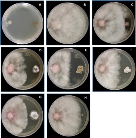
Figure 7. Dual confrontation with Fusarium isolate 1. A) Distribution of <em>Fusarium</em> discs. B) Growth of the control colony of <em>Fusarium</em> isolate 1. C) <em>Fusarium</em> isolate 1 and morphotype M1B M1. D) <em>Fusarium</em> isolate 1 and morphotype M1B M4. G) <em>Fusarium</em> isolate 1 and morphotype M2B M60. H) <em>Fusarium</em> isolate 1 and morphotype M2B M74. I) Mycelial growth of control <em>Fusarium</em> isolate 1.
Figure 7. Dual confrontation with Fusarium isolate 1. A) Distribution of Fusarium discs. B) Growth of the control colony of Fusarium isolate 1. C) Fusarium isolate 1 and morphotype M1B M1. D) Fusarium isolate 1 and morphotype M1B M4. G) Fusarium isolate 1 and morphotype M2B M60. H) Fusarium isolate 1 and morphotype M2B M74. I) Mycelial growth of control Fusarium isolate 1.