RMF Logo

Article filters

Search Papers:

For searching before 2024 see Archive (2000-2023)
  • Vol. 2026, 44(1)
  • Phytopathological Note

Pochonia chlamydosporia var. chlamydosporia on Meloidogyne incognita eggs from Escuinapa, Sinaloa

By Omar Jiménez Pérez, Bernardo Espinosa Palomeque, Augusto Gil Ceballos Ceballos, Francisco Castillo Reyes, Gabriel Gallegos Morales*

*Corresponding Author.

Received: 30/November/2024 – Published: 14/November/2025
DOI: https://doi.org/10.18781/R.MEX.FIT.2024-31

  • PDF
  • HTML
  • XML

Abstract Background/Objective. Chili cultivation is of great importance to Escuinapa, as it is the largest producer of chili in Sinaloa, Mexico. Its production is affected by various pests, such as the Meloidogyne nematode, which can cause up to 50% losses. Control is based on the application of chemical nematicides. Therefore, the need for more sustainable biocontrol alternatives is crucial. The objective of this study was to isolate and identify the nematode and the nematode-parasitic fungus and to evaluate their in vitro parasitism on the eggs of the root-knot nematode of chili plants in Escuinapa, Sinaloa.

Experimental development. Chili roots with nematode galls were collected to isolate and identify the nematode causing the disease and nematophagous fungi. Females and juveniles were identified by perineal sections and morphometry. The nematophagous fungus was identified morphologically and by molecular sequencing (ITS1 and ITS2). The in vitro parasitism of the fungus on Meloidogyne eggs was evaluated at six spore concentrations plus a control. The percentage of parasitism and the LD50 and LD95 were calculated using probit analysis.

Results. Meloidogyne incognita was morphologically identified as the root-knot nematode of chili plants. In turn, Pochonia chlamydosporia var. chlamydosporia was isolated and identified morphologically and molecularly, which showed high percentages of parasitism (98.9%) in vitro on eggs of this nematode over a period of 48 hours.

Conclusion. A strain of Pochonia chlamydosporia was isolated from Meloidogyne incognita that showed high levels of parasitism in eggs (80 to 98%) of this nematode in vitro. In vivo and field trials could confirm its effectiveness as a bionematicide.

Keywords: Alternative, Biocontrol, Nematophagous fungus, In vitro, Root-knot nematode
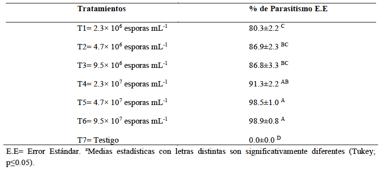
Table 1. Percentage of <em>Pochonia chlamydosporia</em> parasitism on <em>Meloidogyne incognita</em> eggs under <em>in vitro</em> conditions.
Table 1. Percentage of Pochonia chlamydosporia parasitism on Meloidogyne incognita eggs under in vitro conditions.
Table 2. Probabilistic analysis of the concentration of <em>Pochonia chlamydosporia</em> spores on the suppression of <em>Meloidogyne incognita</em> eggs.
Table 2. Probabilistic analysis of the concentration of Pochonia chlamydosporia spores on the suppression of Meloidogyne incognita eggs.
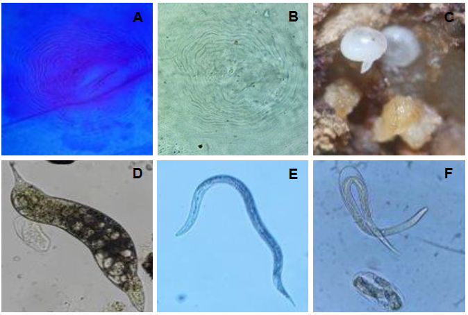
Figure 1. Morphological characteristics of <em>Meloidogyne incognita</em>; A and B) Perianal cut of globose females; C) Globose females; D) Juvenile J4; E) Juvenile stage J2; F) Juvenile stage J2 hatching from the egg.
Figure 1. Morphological characteristics of Meloidogyne incognita; A and B) Perianal cut of globose females; C) Globose females; D) Juvenile J4; E) Juvenile stage J2; F) Juvenile stage J2 hatching from the egg.
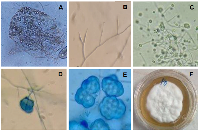
Figure 2. Morphological characteristics of <em>Pochonia chlamydosporia</em> var. <em>chlamydosporia</em>; A) Female globose from which <em>Pochonia</em> was isolated, with the presence of dictyochlamidospores; B) Branched verticillated conidiophores; C) Production of spores in a cluster; D) Formation of a dictyochlamidospore; E) Cluster of dictyochlamidospores; F) Whitish colored mycelium.
Figure 2. Morphological characteristics of Pochonia chlamydosporia var. chlamydosporia; A) Female globose from which Pochonia was isolated, with the presence of dictyochlamidospores; B) Branched verticillated conidiophores; C) Production of spores in a cluster; D) Formation of a dictyochlamidospore; E) Cluster of dictyochlamidospores; F) Whitish colored mycelium.
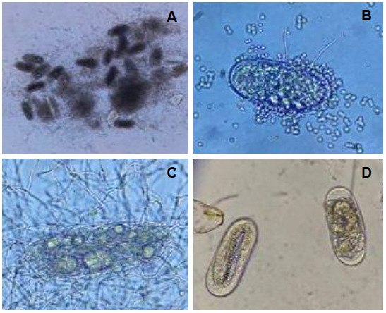
Figure 3. <em>Pochonia chlamydosporia</em> parasitims on <em>Meloidogyne incognita</em> eggs; A) Parasited eggs with a large amount of mycelium; B) Mycelium coming out of the egg and presence of spores on the surface of the egg; C) Parasited egg with mycelium and the presence of dictyochlamidospores; D) Eggs from the control treatment.
Figure 3. Pochonia chlamydosporia parasitims on Meloidogyne incognita eggs; A) Parasited eggs with a large amount of mycelium; B) Mycelium coming out of the egg and presence of spores on the surface of the egg; C) Parasited egg with mycelium and the presence of dictyochlamidospores; D) Eggs from the control treatment.