RMF Logo

Article filters

Search Papers:

For searching before 2024 see Archive (2000-2023)
  • Vol. 2026, 44(1)
  • Scientific Article

Pathogenicity, aggressivity, and inoculum load induced by two ITS-haplotypes of Neopestalotiopsis rosae, isolated from strawberry (Fragaria x ananassa) and blackberry (Rubus sp.)

By Citlalmina Erandy Montes Hernández, Gustavo Mora Aguilera*, María Elizabeth Morales Cortés, Gerardo Acevedo Sánchez, Ángel López Zárate

*Corresponding Author.

Received: 14/June/2024 – Published: 31/December/2025
DOI: https://doi.org/10.18781/R.MEX.FIT.2406-5

  • PDF
  • HTML
  • XML

Abstract Background/Objective. In strawberry (Fragaria x ananassa), the main commercial rosaceous fruit in Mexico, the occurrence of Neopestalotiopsis rosae (Ascomycota: Sporocadaceae) was recently reported in Michoacán. The objective was to analyze, using an etiological-epidemiological approach, the implication of two N. rosae ITS-haplotypes, associated to five isolates from strawberry and blackberry (Rubus sp.) selected for regional prevalence, in the pathogenesis, aggressivity, and inoculum load in strawberry cv. San Andreas.

Materials and Methods. Three isolates of Haplotype 1 / Strawberry (H1): F142P40S, F6P35R, F17P34R, and two of Haplotype 2 (H2) / Blackberry: F142D93R and Z13P27R were cultured in V8 / 25° / 14 d until conidiomata formation, and in PDA / 25° / 7 d to obtain liquid and solid inoculum, respectively. Eight three-month-old strawberry plants cv. San Andreas were evaluated per isolate; two trifoliate leaves / 4 plants were inoculated with 4 mL spray 1 x 10⁶ conidia mL-1, and 4 plants with 5 x 5 mm mycelium PDA-blocks per crown wound. Sterile water was sprayed on 8 control plants. Plants were kept in a semi-covered greenhouse. Starting at 72 hai, every 3 d / 3 months, the following variables were measured: Number of lesions (LN), Lesion diameter (LD), Number of sporulating lesions (EL), Plant height, Crown diameter, and Chlorophyll units SPAD 502 (CU). The incubation period (Pi), latency period (Pl), and generation period (Pg) were estimated for each haplotype. Temperature and RH were recorded every 30 min with HOBO U23. Pathogenicity was validated using Koch's postulates. Isolates H1 and H2 were confirmed culturally by ITS1 / ITS4. ANOVA, in split plots design, and Tukey (p = 0.05) were conducted in RStudio.

Results. The five isolates of N. rosae were more infectious on leaves (p = 0.0001) but exhibited variable and significant aggressivity (p = 0.0001) on LN, EL, and LD, with expression of circular leaf lesions, light brown color, dark edge, and reddish halo on the upper and lower surface. H1 isolates were more pathogenic than H2 (e.g., 3504 vs. 270 LN / plant), with H1 - F142P40S being more aggressive (p = 0.05), with an average LN = 187.5 and EL = 8.45 at 2 and 26 days after inoculation (dai), respectively. In contrast, H2 - F142D93R and H2 – Z13P27R had LN = 20 and 25 at 87 and 95 dai, respectively, with a maximum average range of 0.19 - 0.37 EL > 73 dai. Similarly, in crown, all isolates were pathogenic, but with less aggressivity and no statistical difference. A reddish-dark discoloration and internal necrotic rot was observed in crown and low root density. H1 - F17P34R was conspicuous with a maximum NL = 20 and LE < 0.1 at 85 dai. In leaf, the speed of symptoms expression and sporulation rate, although variable, was higher in isolates H1 vs. H2, determined, respectively, by 2 vs. 4 dai Pi, 14 - 16 (3 d) vs. 33 - 40 (7 d) dai Pl, and 14 - 73 (59 d) vs. 33 - 56 (23 d) dai Pg. The crown infection was slower with 28 d Pi in both haplotypes. Contrasting aggressivity among isolates had no significant effect on plant height and crown diameter, but did have an effect on CU reduction, with a maximum of 14.5 in H1 - F142P40S compared to control. ITS of 33 isolates confirmed the H1 and H2 pathogenicity.

Conclusion. Through significantly contrasting LN, EL, CU, Pi, Pl, and Pg, it was demonstrated the pathogenic variability of two N. rosae ITS-haplotypes, prevalent in Michoacán, obtained from strawberries and blackberries commercial cultivars. A novel mechanistic understanding of pathogenesis was addressed. It is reported for the first time that N. rosae isolates, associated with blackberries, were pathogenic to strawberries but with less aggressivity. Sustainable regional management must consider the variable capacity of the fungus and its interaction with other pathogens with vascular capacity.

Keywords: Berries, Chlorophyll Units, Incubation Period, Latent Period, Physiology
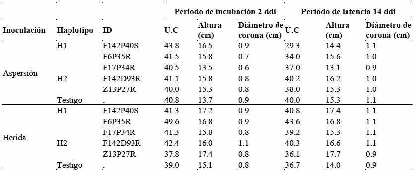
Table 1. Average chlorophyll units (CU), plant height, and crown diameter in strawberry plants cv. San Andreas inoculated by spraying and wounding with five isolates of <em>N. rosae</em>.
Table 1. Average chlorophyll units (CU), plant height, and crown diameter in strawberry plants cv. San Andreas inoculated by spraying and wounding with five isolates of N. rosae.
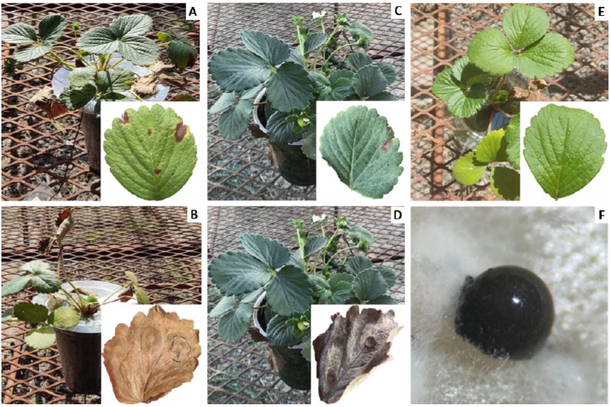
Figure 1. Expression of lesions and sporulating lesions on strawberry leaves cv. San Andreas inoculated by spraying with isolates of <em>N. rosae</em>. <strong>A</strong> and <strong>B</strong>. NL and LE 21 days after inoculation (ddi) with F142P40S. <strong>C</strong> and <strong>D</strong>. Number of lesions (NL) and sporulating lesions (LE) at 77 ddi with isolate Z13P27R <strong>E</strong>. Absolute control, <strong>F</strong>. Conidioma of <em>N. rosae</em> on PDA.
Figure 1. Expression of lesions and sporulating lesions on strawberry leaves cv. San Andreas inoculated by spraying with isolates of N. rosae. A and B. NL and LE 21 days after inoculation (ddi) with F142P40S. C and D. Number of lesions (NL) and sporulating lesions (LE) at 77 ddi with isolate Z13P27R E. Absolute control, F. Conidioma of N. rosae on PDA.
Figure 2. Longitudinal section of crown of strawberry plants cv. San Andreas at 115 ddi inoculated with <em>N. rosae</em>, F142P40S (H1) and F142D93R (H2). In wound inoculation (B), H1 caused total necrosis. The crown shows predominantly light brown to dark reddish-brown color changes. No color change was detected in control plants.
Figure 2. Longitudinal section of crown of strawberry plants cv. San Andreas at 115 ddi inoculated with N. rosae, F142P40S (H1) and F142D93R (H2). In wound inoculation (B), H1 caused total necrosis. The crown shows predominantly light brown to dark reddish-brown color changes. No color change was detected in control plants.
Figure 3. Progress of lesions on leaf blades of strawberry plants cv. San Andreas inoculated by spraying with five isolates of <em>N. rosae</em> corresponding to haplotypes H1 and H2 at 2, 19, 40, 63, and 95 ddi. The dashes represent leaf loss due to total tissue deterioration caused by the aggressiveness associated with each isolate. For example, in F6P35R, leaf loss is reached at 40 dpi.
Figure 3. Progress of lesions on leaf blades of strawberry plants cv. San Andreas inoculated by spraying with five isolates of N. rosae corresponding to haplotypes H1 and H2 at 2, 19, 40, 63, and 95 ddi. The dashes represent leaf loss due to total tissue deterioration caused by the aggressiveness associated with each isolate. For example, in F6P35R, leaf loss is reached at 40 dpi.
Figure 4. Analysis of variance (Tukey, p < 0.05) in split plots, where the large plot is the inoculation method and the small plot is the isolate for the variables number of lesions (<strong>A</strong>) and sporulating lesions (<strong>B</strong>). Different letters indicate significant differences at α = 0.05.
Figure 4. Analysis of variance (Tukey, p < 0.05) in split plots, where the large plot is the inoculation method and the small plot is the isolate for the variables number of lesions (A) and sporulating lesions (B). Different letters indicate significant differences at α = 0.05.
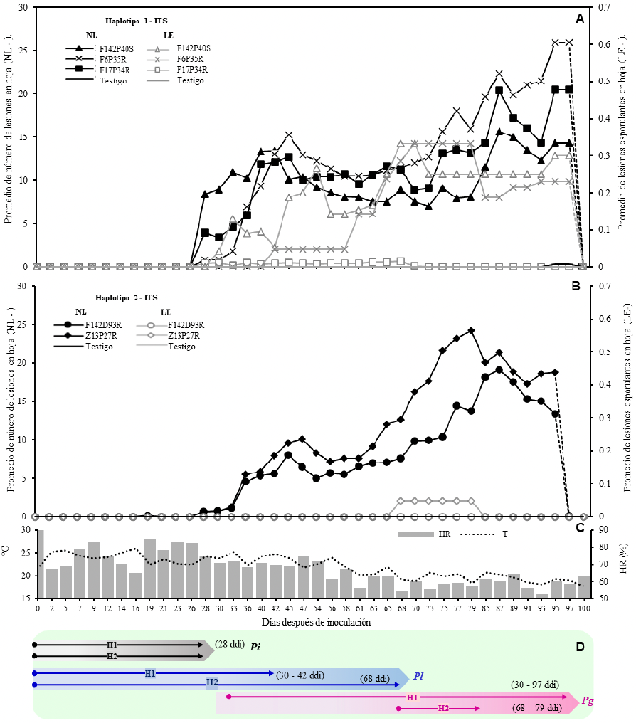
Figure 5. Dynamics of the number of lesions (NL) and sporulating lesions (LE) on strawberry leaves cv. San Andreas up to 95 days after spraying with five isolates of <em>N. rosae</em> obtained from strawberry (F) and blackberry (Z), <strong>A</strong>. Absolute average of NL and LE of three isolates of Haplotype 1 (H1). <strong>B</strong>. Absolute average of NL and LE in two isolates of Haplotype 2 (H2), <strong>C</strong>. Association of RH and T °C with the behavior of the fungus on tissue up to 100 days <strong>D</strong>. Estimation of incubation period (Pi), latency period (Pl), and generation period (Pg) by haplotype of <em>N. rosae</em>.
Figure 5. Dynamics of the number of lesions (NL) and sporulating lesions (LE) on strawberry leaves cv. San Andreas up to 95 days after spraying with five isolates of N. rosae obtained from strawberry (F) and blackberry (Z), A. Absolute average of NL and LE of three isolates of Haplotype 1 (H1). B. Absolute average of NL and LE in two isolates of Haplotype 2 (H2), C. Association of RH and T °C with the behavior of the fungus on tissue up to 100 days D. Estimation of incubation period (Pi), latency period (Pl), and generation period (Pg) by haplotype of N. rosae.
Figure 6. Dynamics of lesions (NL) and sporulating lesions (LE) on leaves up to 95 ddi per wound in the crown with five isolates of <em>N. rosae</em> obtained from strawberries (F) and blackberries (Z), A. Absolute average of NL and LE of three isolates of H1, B. Absolute average of NL and LE of two isolates of H2, C. Association of RH and T °C with the behavior of the fungus on tissue up to 100 days, D. Estimation of incubation period (Pi), latency period (Pl), and generation period (Pg) by <em>N. rosae</em> haplotype.
Figure 6. Dynamics of lesions (NL) and sporulating lesions (LE) on leaves up to 95 ddi per wound in the crown with five isolates of N. rosae obtained from strawberries (F) and blackberries (Z), A. Absolute average of NL and LE of three isolates of H1, B. Absolute average of NL and LE of two isolates of H2, C. Association of RH and T °C with the behavior of the fungus on tissue up to 100 days, D. Estimation of incubation period (Pi), latency period (Pl), and generation period (Pg) by N. rosae haplotype.