RMF Logo

Article filters

Search Papers:

For searching before 2024 see Archive (2000-2023)
  • Vol. 2025, 43(3)
  • Scientific Article

Fatty acid composition of lipopeptides with antifungal activity synthesized by Bacillus amyloliquefaciens

By Nancy Ley López, José Basilio Heredia, Isabel Cruz Lachica, Isidro Márquez Zequera, Raymundo Medina López, Cesar San Martín Hernández, Raymundo Saúl García Estrada*

*Corresponding Author.

Received: 14/May/2025 – Published: 18/July/2025
DOI: https://doi.org/10.18781/R.MEX.FIT.2503-1

  • PDF
  • HTML
  • XML

Abstract Background/Objective. Some Bacillus species synthesize biologically active lipopeptides containing fatty acids of varying length. Fatty acid composition is key to their potential activity as surfactants. The objective was to determine the composition and quantification of fatty acids in the structures of lipopeptides synthesized by Bacillus amyloliquefaciens KX953161.1 and to observe the effect of lipopeptides on the mycelial growth of Fusarium oxysporum MG557870 and Sclerotium rolfsii OM510466.

Materials and Methods. The lipid profile of the extract with lipopeptides synthesized by B. amyloliquefaciens KX953161.1 was performed by gas chromatography with a flame ionization detector (GC-FID). The quantification of the detected fatty acids was performed using the area standardization method, and the fat determination was carried out using the 920.39 AOAC method. In addition, scanning electron microscopy (SEM) was used to observe the inhibitory effect of lipopeptides on the mycelial growth of F. oxysporum MG557870 and S. rolfsii OM510466. The data obtained (determination of fatty acids and MIC) were analyzed by analysis of variance (ANOVA), and the comparison of means was performed using the Tukey test (p≤0.05).

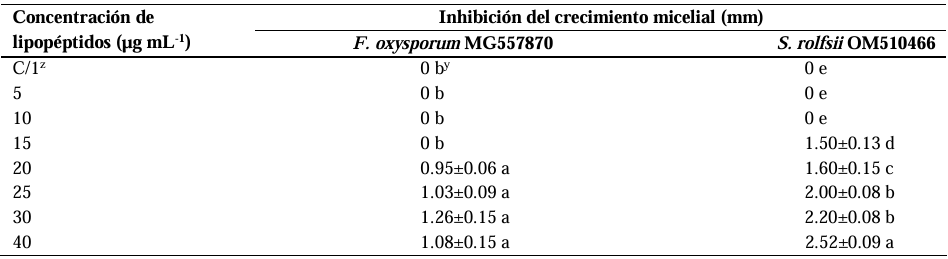
Results. Variability was observed in C13 to C18 β-hydroxylated fatty acids. The C14 β hydroxylated fatty acid (tetradecanoic acid) had the highest content, with 8.254 ± 0.031 ng mg-1 of sample, followed by the C13 β-hydroxylated fatty acids (tridecanoic acid) and C16 fatty acids (palmitic acid), with 4.304 ± 0.064 and 4.100 ± 0.120 ng mg-1, respectively. The minimum inhibitory concentration (MIC) of the lipopeptides on F. oxysporum MG557870 and S. rolfsii OM510466 was 20 and 15 µg mL-1. Damage to fungal cells was evident in their structure and morphology.

Conclusion. Lipopeptide synthesis by B. amyloliquefaciens KX953161.1 exhibits variation in the length of the fatty acid chains, which are primarily composed of C14 (tetradecanoic) fatty acids. This variability could be related to the biological activity of the lipopeptides, and the C14 (tetradecanoic) fatty acid could be important in their antagonism against phytopathogenic fungi.

Keywords: Surfactant activity, β-hydroxylated, Tetradecanoic, Fusarium oxysporum, Sclerotium rolfsii

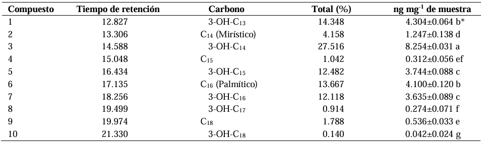
Table 1. Conditions of injection by Gas Chromatography (GC).
Table 2. Profile of fatty acids of the lipopeptides biosynthesized by <em>Bacillus amyloliquefaciens</em> KX953161.1, identified using GC -MS.
Table 2. Profile of fatty acids of the lipopeptides biosynthesized by Bacillus amyloliquefaciens KX953161.1, identified using GC -MS.
Table 3. Antifungal effect of the lipopeptides synthesized by <em>B. amyloliquefaciens</em> KX953161.1 with the CMI on the mycelial growth of <em>F. oxysporum</em> MG557870 and <em>S. rolfsii</em> OM510466.
Table 3. Antifungal effect of the lipopeptides synthesized by B. amyloliquefaciens KX953161.1 with the CMI on the mycelial growth of F. oxysporum MG557870 and S. rolfsii OM510466.
Figure 1. Chromatogram of the lipid profile of the lipopeptides synthesized by <em>Bacillus amyloliquefaciens</em> KX953161.1, analyzed by CG-MS.
Figure 1. Chromatogram of the lipid profile of the lipopeptides synthesized by Bacillus amyloliquefaciens KX953161.1, analyzed by CG-MS.
Figure 2. Mass spectrum of fatty acid C<sub>14</sub> (Tetradecanoic) of the extract with lipopeptides synthesized by <em>B. amyloliquefaciens</em> KX953161.1.
Figure 2. Mass spectrum of fatty acid C14 (Tetradecanoic) of the extract with lipopeptides synthesized by B. amyloliquefaciens KX953161.1.
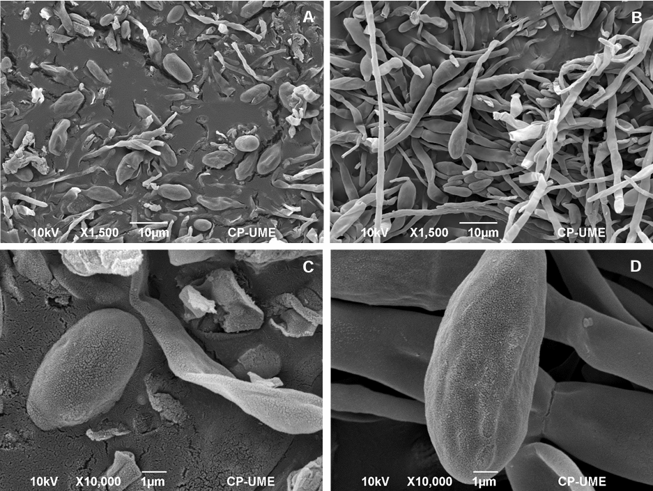
Figure 3. Micrographs (MEB) of spores and hyphae from <em>Fusarium oxysporum</em> MG557870 after the treatment with lipopeptides synthesized by <em>Bacillus amyloliquefaciens</em> KX953161.1. A and C: spores and hyphae treated with lipopeptides (1,500x, bar= 10 µm and 10,000x, bar= 1 µm, respectively); B and D: spores and hyphae without lipopeptides (1,500x, bar= 10 µm y 10,000x, bar= 1 µm, respectively).
Figure 3. Micrographs (MEB) of spores and hyphae from Fusarium oxysporum MG557870 after the treatment with lipopeptides synthesized by Bacillus amyloliquefaciens KX953161.1. A and C: spores and hyphae treated with lipopeptides (1,500x, bar= 10 µm and 10,000x, bar= 1 µm, respectively); B and D: spores and hyphae without lipopeptides (1,500x, bar= 10 µm y 10,000x, bar= 1 µm, respectively).
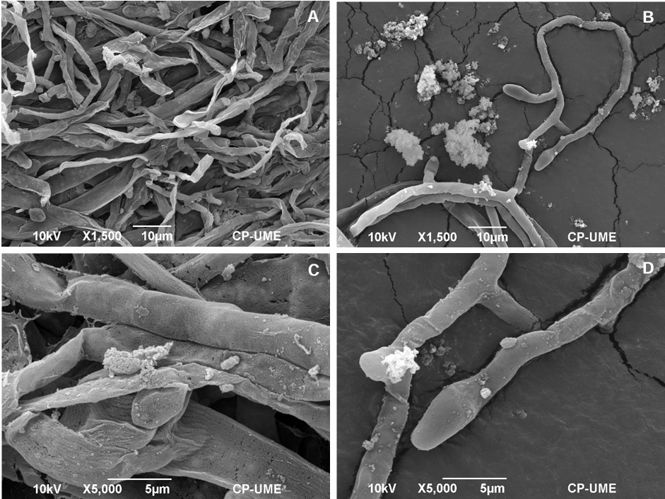
Figure 4. Micrographs (MEB) of mycelia from <em>Sclerotium rolfsii</em> OM510466 after the treatment with lipopeptides synthesized by <em>Bacillus amyloliquefaciens</em> KX953161.1. A and C: mycelia treated with lipopeptides (1,500x, bar= 10 µm and 5,000x, bar= 5 µm, respectively); B y D: micelium without lipopeptides (1,500x, bar= 10 µm y 5,000x, bar= 5 µm, respectively).
Figure 4. Micrographs (MEB) of mycelia from Sclerotium rolfsii OM510466 after the treatment with lipopeptides synthesized by Bacillus amyloliquefaciens KX953161.1. A and C: mycelia treated with lipopeptides (1,500x, bar= 10 µm and 5,000x, bar= 5 µm, respectively); B y D: micelium without lipopeptides (1,500x, bar= 10 µm y 5,000x, bar= 5 µm, respectively).