Click on the cite to copy
-
Or Copy link


Article filters
Search Papers:
Potential antagonist of native rhizobacteria of Parkinsonia aculeata on Fusarium spp. associated with native maize varieties from Bajío, Mexico
By Leandris Argentel Martínez, Ofelda Peñuelas Rubio*, Francisco Cervantes Ortíz, Joe Luis Arias Moscoso, Francisco Cadena Cadena, Pamela Romo Rodríguez, Lorenzo Pérez López, Rosario Alicia Fierro Coronado
*Corresponding Author.
Received: 15/January/2026 – Published:
29/April/2026
DOI:
https://doi.org/10.18781/R.MEX.FIT.2601-2
-
Or Copy link
Abstract Background/Objective. The genus Fusarium comprises some of the most important phytopathogenic fungi affecting maize crops. Fusarium cause diseases such as root, stalk, and ear rot, which negatively impact the crop's agricultural productivity. In the search for agrobiotechnological alternatives for controlling this fungus, this study was aimed to evaluate the antagonistic potential of Parkinsonia aculeata rhizobacteria against Fusarium spp. strains associated with native maize varieties from the Bajío region of Mexico.
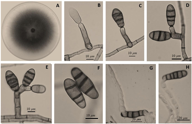
Experimental development. Two strains of Fusarium spp. (MC-03 and MC-05), isolated from roots of native maize varieties from the Bajío region of Mexico, exhibiting Fusarium wilt symptoms, were used. These fungi were tested in vitro against nine rhizobacteria of P. aculeata: Enterobacter cloacae (BA1), Priestia megaterium (BA4 and BA-7B), Sinomonas halotolerans (BA10-B), Staphylococcus warneri (BP5), P. endophytica (BP6), Bacillus subtilis (TP1 and TP2), and S. hominis (TM6). Microscopic and macroscopic characterization of the fungal strains and biochemical analysis of the rhizobacteria were performed. Radial growth inhibition of the fungal isolates was determined by triplicate dual fungus-rhizobacteria confrontations. A completely randomized design was used, analyzing the data obtained in the STATISTICA software using an ANOVA based on a linear model of fixed effects and a mean comparison test by DMS (p>0.05).
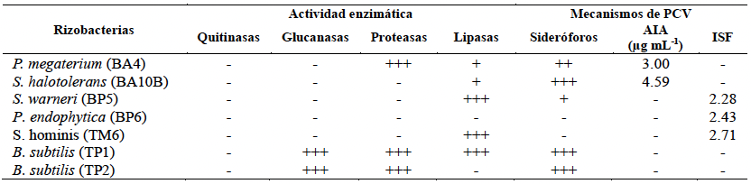
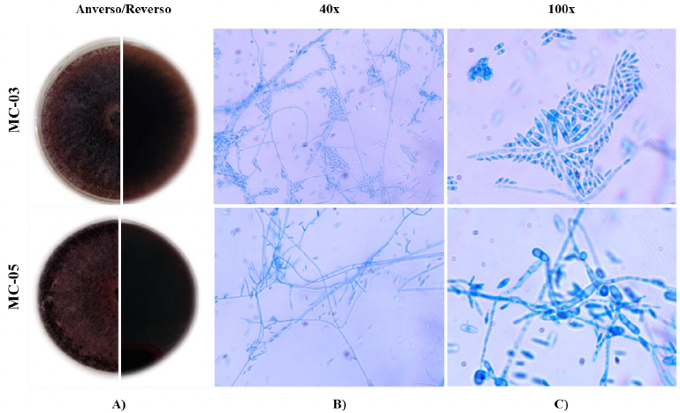
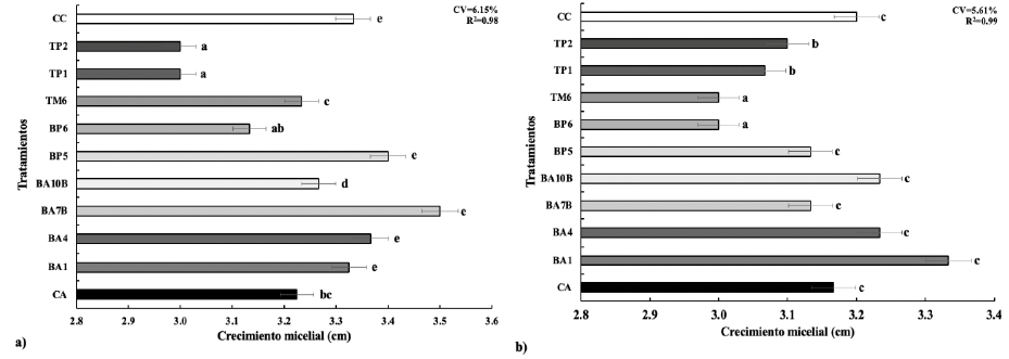
Results. With the exception of P. endophytica (BP6), all rhizobacteria exhibited enzymatic activity related to fungal antagonism mechanisms. B. subtilis (TP1) produced glucanases, lipases, and proteases. Microscopic and macroscopic characterization of the fungal strains indicated that they belong to Fusarium spp. In the rhizobacteria-MC-03 confrontation, the B. subtilis bacterial strains (TP1 and TP2) were statistically similar and achieved the greatest inhibition of mycelial growth (23%). With the fungal strain MC-05, P. endophytica (BP6) and S. hominis (TM6) were statistically superior to the other rhizobacteria in inhibiting mycelial growth (17%), followed by the two B. subtilis strains (TP1 and TP2), which inhibited 10% and 17%, respectively.
Conclusion. There was a significant variability in the mycelial growth response of the fungi to rhizobacteria. B. subtilis (TP1 and TP2), S. hominis (TM6), and P. endophytica (BP6) exhibited an antagonistic effect, inhibiting the mycelial growth of Fusarium spp. strains by up to 23% compared to the absolute and commercial controls. This study establishes the preliminary scientific basis for obtaining a biofungicide with specific inhibitory capabilities against the fungi studied.
Keywords: Bacillus subtilis, Phytopathogenic fungi, Priestia endophytica, Staphylococcus hominis, Zea mays