RMF Logo

Article filters

Search Papers:

For searching before 2024 see Archive (2000-2023)
  • Vol. 2026, 44(3)
  • Phytopathological Note

In vitro antifungal activity and sensitivity of Neopestalotiopsis rosae isolates to Baccharis salicifolia and Fouquieria splendens extracts

By Jesús Eduardo Ramírez Méndez, Meralida Bartolón Pérez, Francisco Daniel Hernández Castillo, Yisa María Ochoa Fuentes, Laura Castro Rosalez, Marco Antonio Tucuch Pérez*

*Corresponding Author.

Received: 19/January/2026 – Published: 13/July/2026
DOI: https://doi.org/10.18781/R.MEX.FIT.2601-3

  • PDF
  • HTML
  • XML

Abstract Background/Objective. Neopestalotiopsis rosae is the causal agent of crown and root rot in strawberry, resulting in significant yield losses. The aim of this study was to evaluate the sensitivity of four N. rosae isolates (466, 323, FREC, and 372) to plant extracts (PEs) obtained from Baccharis salicifolia and Fouquieria splendens, and to determine their in vitro antifungal activity.

Experimental development. A qualitative phytochemical screening was performed to identify the major bioactive compounds present in the PEs. Antifungal activity was evaluated using in vitro microdilution assays at concentrations ranging from 3.9 to 2 000 mg L⁻¹ (10 concentrations). The 50 and 90% inhibitory concentrations (IC₅₀ and IC₉₀) were estimated, and the data were analyzed using analysis of variance (ANOVA) followed by Tukey's multiple comparison test (p ≤ 0.05). The effects of the PEs on fungal cellular structures were also assessed.

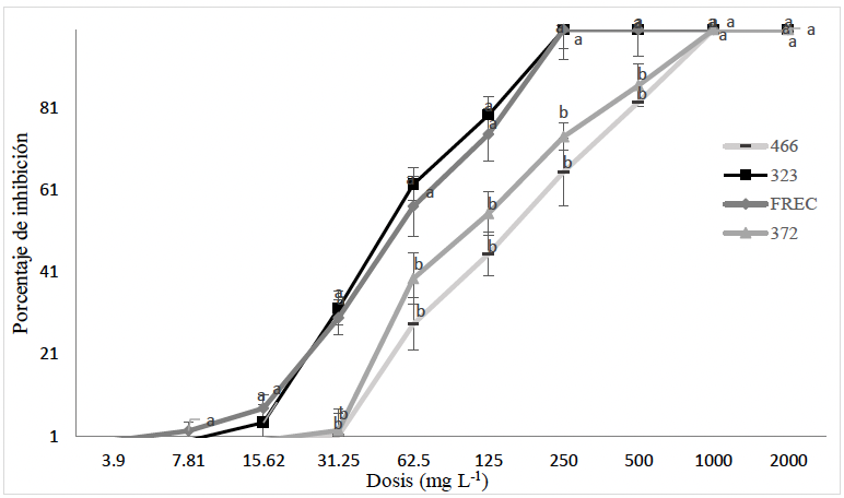
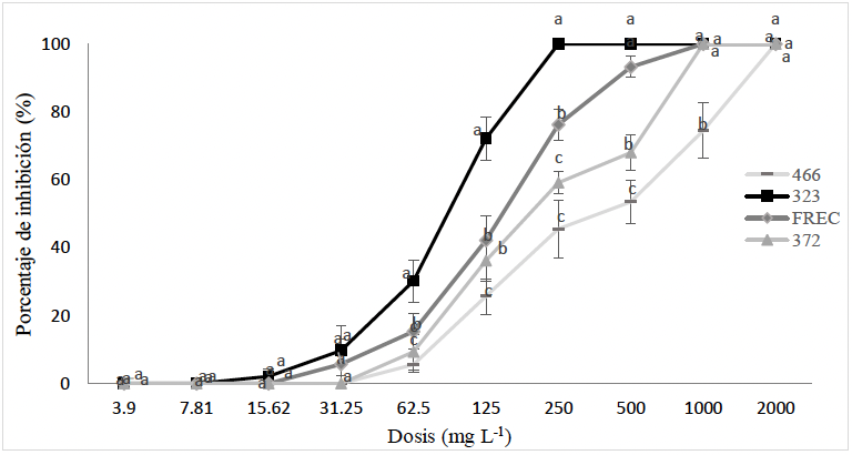
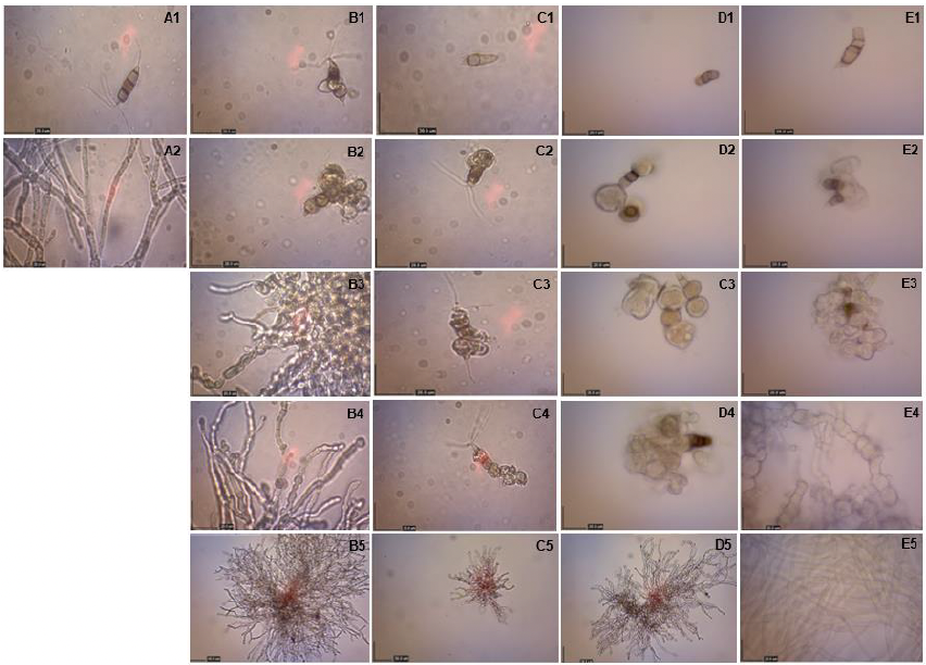
Results. Phytochemical screening revealed the presence of phenolic compounds, saponins, and tannins in both plant extracts. Among the N. rosae isolates, isolates 323 and FREC were the most sensitive, exhibiting complete inhibition with both PEs at 250 mg L⁻¹. In contrast, isolates 372 and 466 required higher concentrations to achieve complete inhibition. B. salicifolia was more effective against isolates 466 and 372, achieving complete inhibition at 1 000 mg L⁻¹, whereas F. splendens required 2 000 mg L⁻¹ to completely inhibit isolate 466. Structural analyses revealed that isolate 323 was most susceptible to extract-induced damage at 250 mg L⁻¹ and above, as evidenced by inhibition of conidial germination and severe morphological alterations, including cellular collapse and dehydration. Isolates FREC and 372 exhibited mycelial thickening at 500 mg L⁻¹, whereas isolate 466 was the least sensitive, showing no evident structural damage even at this concentration.

Conclusions. Plant extracts from B. salicifolia and F. splendens contain bioactive phytochemicals with antifungal activity against N. rosae. Isolates 323 and FREC were the most sensitive, exhibiting 100% inhibition and pronounced structural damage at 250 mg L⁻¹, highlighting the potential of these extracts as biorational alternatives for the management of N. rosae. Nevertheless, further greenhouse and field studies are required to validate their efficacy under commercial production conditions.

Keywords: Biorational control, Phytochemicals, Antifungal activity, Phytopathogenic, Sustainable agriculture
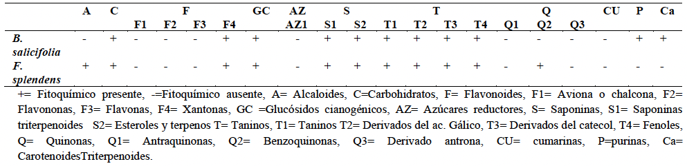
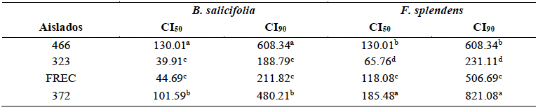
Table 1. Identification of phytochemicals present in methanolic extracts of <em>Baccharis salicifolia</em> and <em>Fouquieria splendens</em> using colorimetric assays.
Table 1. Identification of phytochemicals present in methanolic extracts of Baccharis salicifolia and Fouquieria splendens using colorimetric assays.
Table 2. Fifty percent and ninety percent inhibitory concentrations (IC₅₀ and IC₉₀) of <em>Neopestalotiopsis rosae</em> isolates treated with <em>Baccharis salicifolia</em> and <em>Fouquieria splendens</em> extracts.
Table 2. Fifty percent and ninety percent inhibitory concentrations (IC₅₀ and IC₉₀) of Neopestalotiopsis rosae isolates treated with Baccharis salicifolia and Fouquieria splendens extracts.
Figure 1. Inhibition percentage of <em>Neopestalotiopsis rosae</em> isolates treated with <em>Baccharis salicifolia</em> extract. *Data followed by the same letter are not significantly different
Figure 1. Inhibition percentage of Neopestalotiopsis rosae isolates treated with Baccharis salicifolia extract. *Data followed by the same letter are not significantly different
Figure 2. Inhibition percentage of <em>Neopestalotiopsis rosae</em> isolates treated with <em>Fouquieria splendens</em> extract. *Data followed by the same letter are not significantly different.
Figure 2. Inhibition percentage of Neopestalotiopsis rosae isolates treated with Fouquieria splendens extract. *Data followed by the same letter are not significantly different.
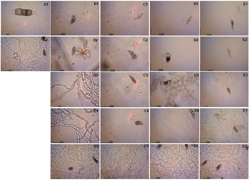
Figure 3. Effect of <em>Baccharis salicifolia</em> extract on the fungal structures of <em>Neopestalotiopsis rosae</em> isolates. A1–A2: untreated control. B1–B5: isolate 466 (2 000, 1 000, 500, 250, and 125 mg L⁻¹); C1–C5: isolate 323 (2 000, 1 000, 500, 250, and 125 mg L⁻¹); D1–D5: isolate FREC (2 000, 1 000, 500, 250, and 125 mg L⁻¹); E1–E5: isolate 372 (2 000, 1 000, 500, 250, and 125 mg L⁻¹). Microscopic observations were performed at 10× and 40× magnification.
Figure 3. Effect of Baccharis salicifolia extract on the fungal structures of Neopestalotiopsis rosae isolates. A1–A2: untreated control. B1–B5: isolate 466 (2 000, 1 000, 500, 250, and 125 mg L⁻¹); C1–C5: isolate 323 (2 000, 1 000, 500, 250, and 125 mg L⁻¹); D1–D5: isolate FREC (2 000, 1 000, 500, 250, and 125 mg L⁻¹); E1–E5: isolate 372 (2 000, 1 000, 500, 250, and 125 mg L⁻¹). Microscopic observations were performed at 10× and 40× magnification.
Figure 4. Effect of <em>Fouquieria splendens</em> extract on the fungal structures of <em>Neopestalotiopsis rosae</em> isolates. A1–A2: untreated control. B1–B5: isolate 466 (2 000, 1 000, 500, 250, and 125 mg L⁻¹); C1–C5: isolate 323 (2 000, 1 000, 500, 250, and 125 mg L⁻¹); D1–D5: isolate FREC (2 000, 1 000, 500, 250, and 125 mg L⁻¹); E1–E5: isolate 372 (2 000, 1 000, 500, 250, and 125 mg L⁻¹). Microscopic observations were performed using 10×, 40×, and 100× objective lenses
Figure 4. Effect of Fouquieria splendens extract on the fungal structures of Neopestalotiopsis rosae isolates. A1–A2: untreated control. B1–B5: isolate 466 (2 000, 1 000, 500, 250, and 125 mg L⁻¹); C1–C5: isolate 323 (2 000, 1 000, 500, 250, and 125 mg L⁻¹); D1–D5: isolate FREC (2 000, 1 000, 500, 250, and 125 mg L⁻¹); E1–E5: isolate 372 (2 000, 1 000, 500, 250, and 125 mg L⁻¹). Microscopic observations were performed using 10×, 40×, and 100× objective lenses