RMF Logo

Article filters

Search Papers:

For searching before 2024 see Archive (2000-2023)
  • Vol. 2026, 44(2)
  • Phytopathological Note

Detection of a tentative alphanucleorhabdovirus infecting Carica papaya in Costa Rica

By Laura Garita Salazar, William Villalobos Muller, Mauricio Montero Astúa, Antonio Bogantes Arias, Teresita Coto Morales, Izayana Sandoval Carvajal, Lisela Moreira Carmona*

*Corresponding Author.

Received: 11/February/2026 – Published: 29/April/2026
DOI: https://doi.org/10.18781/R.MEX.FIT.2602-3

  • PDF
  • HTML
  • XML

Abstract Background/Objective. Foliar chlorosis, short internodes, and curved petioles with purple streaks were observed in papaya (Carica papaya) crops (North and Atlantic regions) in Costa Rica since 2014. Identification of a putative plant virus associated with these symptoms was the aim of this research.

Experimental development. Plant material was tested by ELISA (four plant viruses and potyviruses group), transmission electron microscopy (TEM), RT-PCR (degenerate primers for plant viruses), sequencing and phylogenetics.

Results. All ELISA tests resulted negative. Bullet-shaped particles inside nuclei, and reticulum endoplasmic were only observed by TEM in symptomatic plants. Amplicons of 900 bp were consistently obtained from symptomatic samples using degenerate primers for plant rhabdoviruses. Nucleotide sequences showed 95.6 and 96.8% similarity to a putative papaya alphanucleorhabdovirus (Alphanucleorhabdovirus, Rhabdoviridae).

Conclusion. This is the first report of a putative alphanucleorhabdovirus associated with symptomatic papaya plants showing streaked petiole ("pecíolo rayado") disease in Costa Rica, but Koch's postulates must be fulfilled and vector identified.

Keywords: Plant rhabdoviruses, "Streaked petiole" disease, Papaya necrosis apical
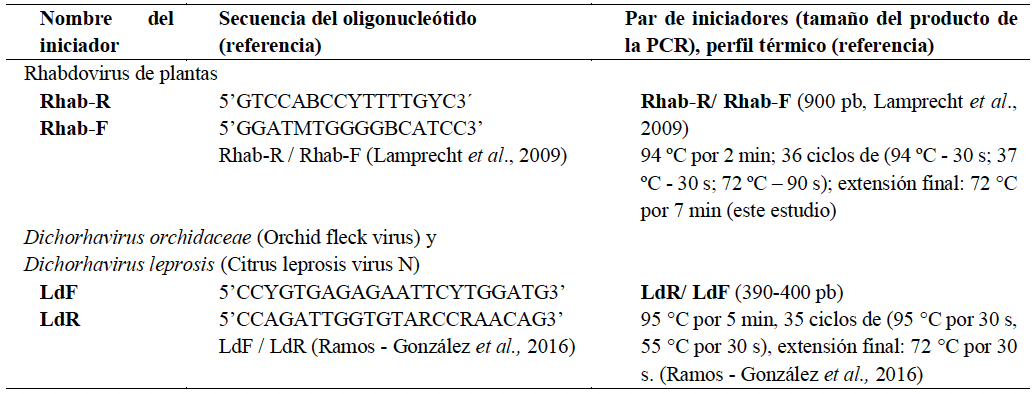
Table 1. Oligonucleotides (primers) and thermocycling profiles used to detect RNA plant viruses in <em>Carica papaya</em> plants analyzed in this study.
Table 1. Oligonucleotides (primers) and thermocycling profiles used to detect RNA plant viruses in Carica papaya plants analyzed in this study.
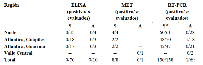
Table 2. Summary of tests performed and results obtained from foliar samples of symptomatic and asymptomatic <em>Carica papaya</em> plants collected in Costa Rica to determine whether any plant virus is associated with
Table 2. Summary of tests performed and results obtained from foliar samples of symptomatic and asymptomatic Carica papaya plants collected in Costa Rica to determine whether any plant virus is associated with "streaked petiole" disease.
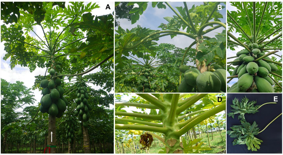
Figure 1. Morphological alterations observed in <em>Carica papaya</em> plants with
Figure 1. Morphological alterations observed in Carica papaya plants with "streaked petiole" disease in Costa Rica. A) A symptomatic plant (arrow) exhibiting downward-curved petioles, leaf reduction in the apical meristem, and cessation of fruit set. Adjacent (slightly behind) two healthy plants (right) showing prolific fruit development and typical foliar architecture; B) Detailed view of upper part of a symptomatic plant showing leaf reduction in the apical meristem, leaf petiole curved, and poor fruit setting, behind (left) a healthy plant with normal fruit production; C) Detailed view of upper part of a healthy plant; D) Petioles of a diseased plant displaying purple-colored streaks; E) Comparison between petioles from symptomatic (top) and healthy (bottom) plants.
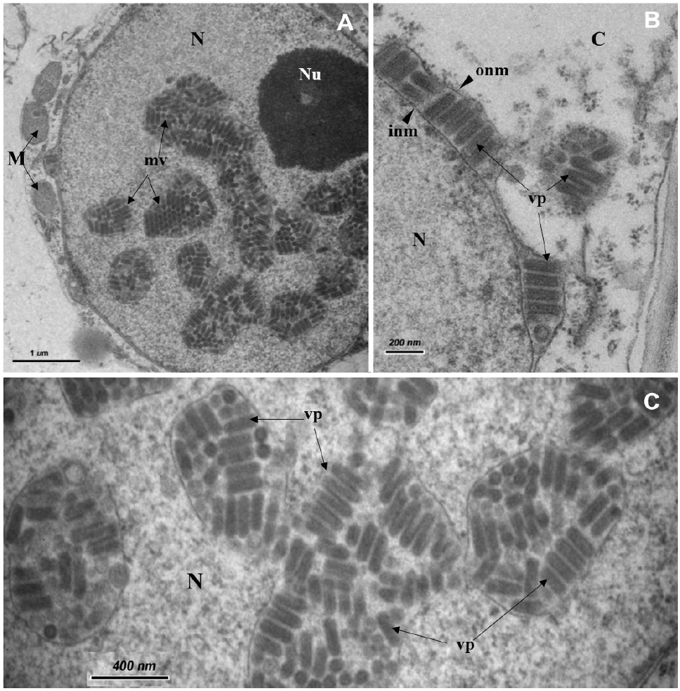
Figure 2. Nucleus of a leaf parenchyma cell of <em>Carica papaya</em> symptomatic plant with "streaked petiole" disease observed with transmission electron microscopy.<strong> A</strong>) Bullet-shaped particles inside membranous vesicles in the nucleus; <strong>B</strong>) Detail of particles seen into the perinuclear space; <strong>C</strong>) Detail of viral particles into the packages scattered in the nucleus. C= cytoplasm, M= mitochondria, N= nucleus, Nu= nucleolus, inm= inner nuclear membrane, mv= membranous vesicle filled of viral particles, onm= outer nuclear membrane, vp= viral particles.
Figure 2. Nucleus of a leaf parenchyma cell of Carica papaya symptomatic plant with "streaked petiole" disease observed with transmission electron microscopy. A) Bullet-shaped particles inside membranous vesicles in the nucleus; B) Detail of particles seen into the perinuclear space; C) Detail of viral particles into the packages scattered in the nucleus. C= cytoplasm, M= mitochondria, N= nucleus, Nu= nucleolus, inm= inner nuclear membrane, mv= membranous vesicle filled of viral particles, onm= outer nuclear membrane, vp= viral particles.
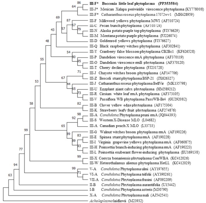
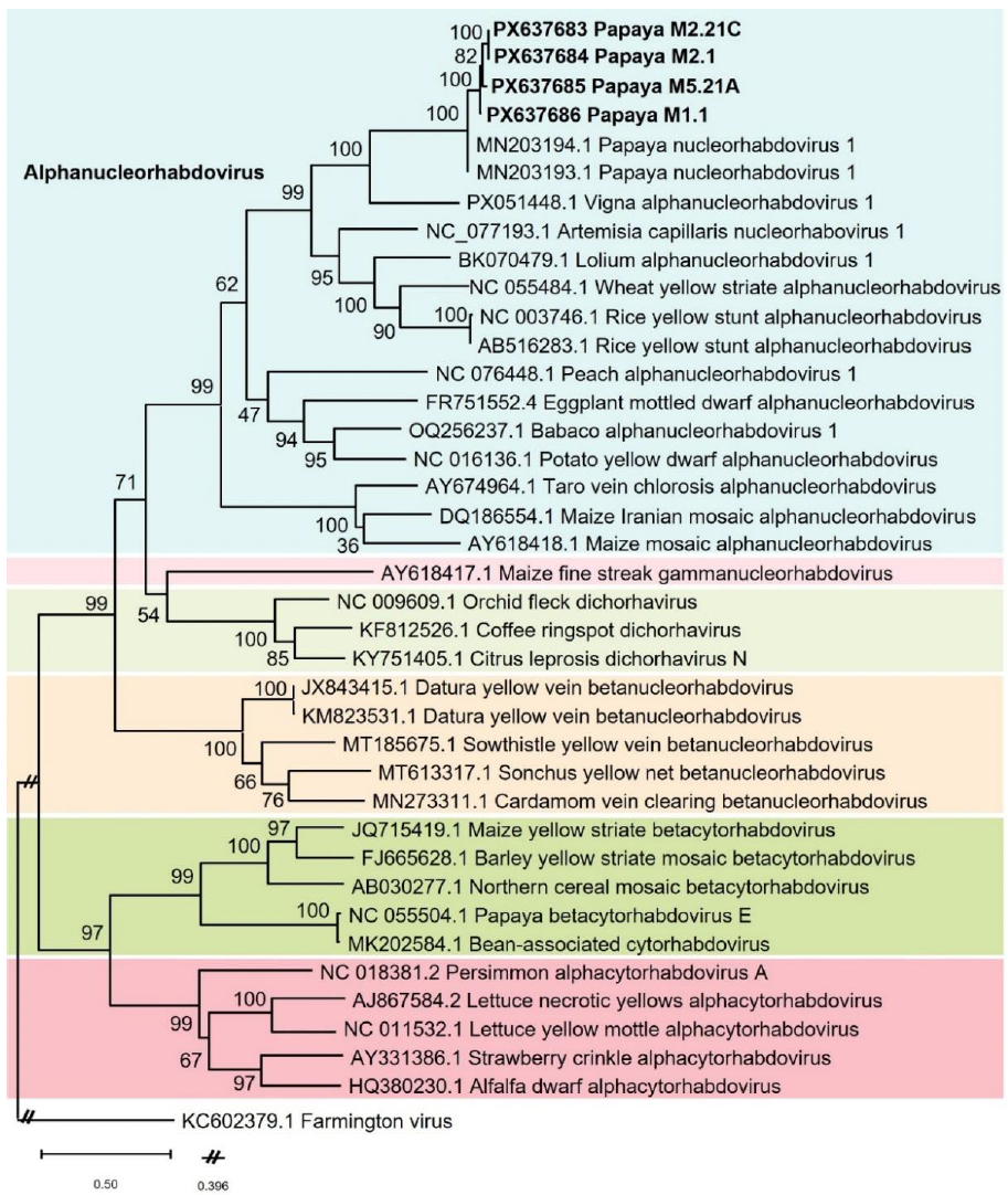
Figure 3. Phylogenetic tree constructed with partial sequences (900 nt) of the viral RNA-dependent RNA polymerase (L gene) of a putative alphanucleorhabdovirus obtained from <em>Carica papaya</em> of Costa Rica (GB Acc. No. PX637683 to PX637686) with 31 different plant rhabdoviruses (34 sequences) and Farmington virus (GB Acc. No. KC602379) as outgroup, all of them retrieved from GenBank. These were aligned with ClustalW algorithm using BioEdit v.7.7.1, and phylogeny inferred with the maximum likelihood method in Mega 12 (General Time Reversible model with a gamma distributed rate of substitution with invariant sites) and a bootstrap of 1 000 replicates.
Figure 3. Phylogenetic tree constructed with partial sequences (900 nt) of the viral RNA-dependent RNA polymerase (L gene) of a putative alphanucleorhabdovirus obtained from Carica papaya of Costa Rica (GB Acc. No. PX637683 to PX637686) with 31 different plant rhabdoviruses (34 sequences) and Farmington virus (GB Acc. No. KC602379) as outgroup, all of them retrieved from GenBank. These were aligned with ClustalW algorithm using BioEdit v.7.7.1, and phylogeny inferred with the maximum likelihood method in Mega 12 (General Time Reversible model with a gamma distributed rate of substitution with invariant sites) and a bootstrap of 1 000 replicates.