RMF Logo

Article filters

Search Papers:

For searching before 2024 see Archive (2000-2023)
  • Vol. 2026, 44(3)
  • Scientific Article

Emerging disease in melon crop associated with a mixed infection by three whitefly-transmitted viruses in La Comarca Lagunera, Mexico

By Ángela Paulina Arce Leal, Edgar Antonio Rodríguez Negrete, Manuel Arturo Méndez De León, Enrique Alejandro Guevara Rivera, Norma Elena Leyva López, Jesús Méndez Lozano*

*Corresponding Author.

Received: 14/April/2026 – Published: 05/August/2026
DOI: https://doi.org/10.18781/R.MEX.FIT.2604-3

  • PDF
  • HTML
  • XML
  • Supplementary Material

Abstract Background/Objective. Cucurbits are an economic pillar of global and national agriculture. However, their productivity is threatened by the increasing incidence of viral plant diseases. Consequently, coinfections with members of the Begomovirus and Crinivirus genera have been associated with exacerbated symptoms and high incidence rates in crops, leading to emerging disease patterns. The aim of this study was to identify and molecularly characterize the begomoviruses and criniviruses associated with an emerging disease in melon crops in a municipality of the Comarca Lagunera region, Mexico.

Materials and Methods. A targeted sampling of nine melon plants exhibiting symptoms such as severe interveinal chlorosis, mottling, yellow mosaic patterns, and fruit deformation was carried out. For viral detection, DNA extraction was performed using 3% CTAB and RNA extraction using the TRIzol reagent. Begomoviruses and criniviruses were detected using PCR and RT-PCR with specific primers. Complete begomovirus genomes were enriched by rolling circle amplification (RCA). Amplicons derived from Open Reading Frame (ORF) of the capsid protein (CP) of criniviruses were generated using RT-PCR. Subsequently, the genomes and CP amplicons were cloned and sequenced for phylogenetic and evolutionary analysis.

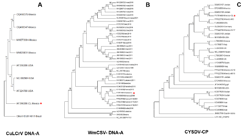
Results. Molecular identification revealed the prevalence of mixed infections in seven of nine samples, comprised of two begomovirus species, cucurbit leaf crumple virus (CuLCrV, Begomovirus cucurbitae) and watermelon chlorotic stunt virus (WmCSV, Begomovirus citrulli), and the crinivirus cucurbit yellow stunting disorder virus (CYSDV, Crinivirus cucurbitae). Genomic analyses confirmed that CuLCrV and WmCSV possess genomic structure typical of begomoviruses. Significant divergence was observed at specific loci (CuLCrV C4 gene, WmCSV NSP gene, and DNA-B intergenic regions of both species), suggesting their potential involvement in evolutionary adaptation processes. Phylogenetically, although the CuLCrV isolate showed close relationship to isolates from Arizona and Sonora, it was placed in a clade distinct from those previously reported in the region, indicating an independent evolutionary course. Furthermore, phylogenetic analysis based on the nucleotide sequence of the CYSDV capsid protein (CP) gene revealed a close relationship with isolates previously reported in Jordan and Mexico.

Conclusions. This study represents the first report in Mexico of a disease associated with WmCSV, CuLCrV, and CYSDV coinfection in cucurbits. The findings amphasize the complexity of emerging diseases and highlight the importance of strengthening molecular epidemiological surveillance to mitigate the impact of these mixed infections on Mexican agriculture.

Keywords: Cucurbits, Begomovirus, Crinivirus, Mixed infections
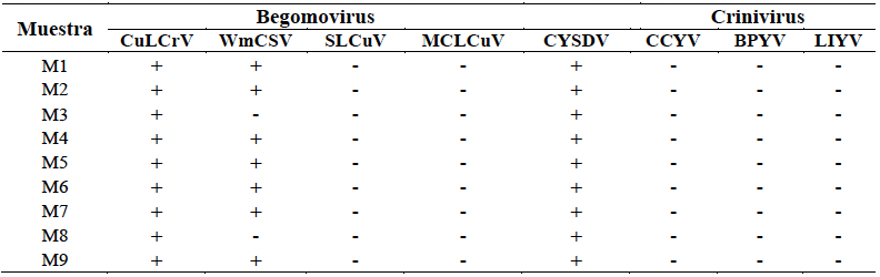
Table 1. Molecular detection of begomoviruses and criniviruses in melon leaf tissue. Nine samples were analyzed by PCR for the detection of begomoviruses: cucurbit leaf crumple virus (CuLCrV), watermelon chlorotic stunt virus (WmCSV), squash leaf curl virus (SLCuV), and melon chlorotic leaf curl virus (MCLCuV), and RT-PCR for the criniviruses: cucurbit yellow stunting disorder virus (CYSDV), cucurbit chlorotic yellows virus (CCYV), beet pseudo yellows virus (BPYV), and lettuce infectious virus (LIVY). (+) positive detection, (-) negative detection.
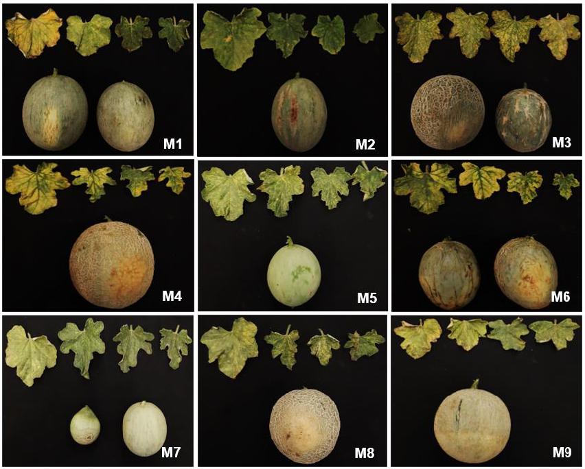
Figure 1. Melon plants and fruits showing viral symptoms collected in the La Comarca Lagunera (CL) region. A total of nine samples (M1-M9) of melon with viral symptoms were collected in the municipality of Tlahualilo. Symptoms observed in the leaf tissue were severe interveinal chlorosis, mottling, and yellow mosaic, while in the fruits, chlorosis, deformation, and cracking.
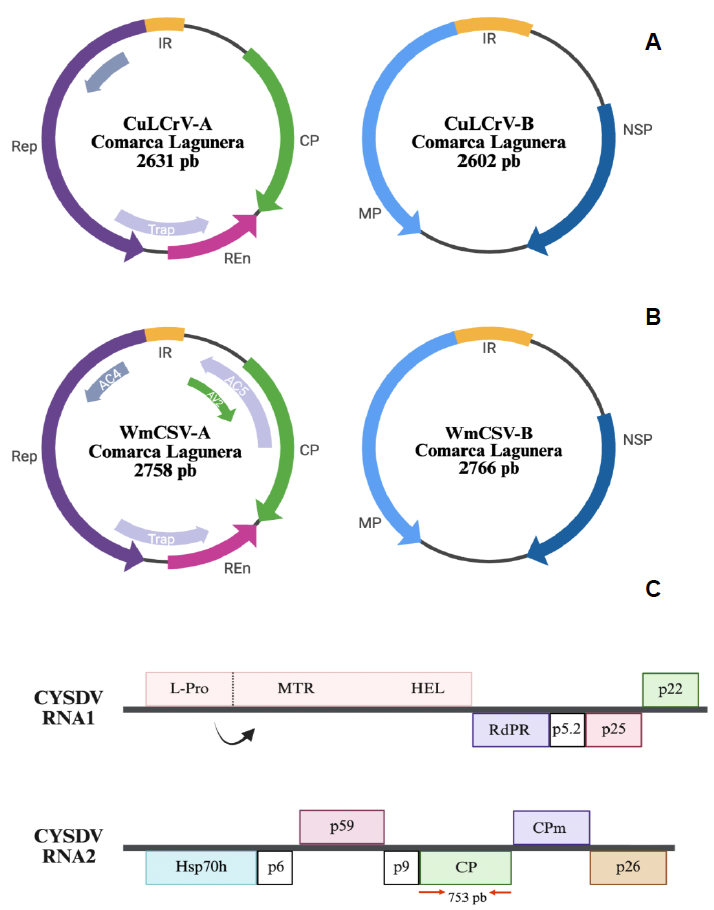
Figure 2. Complete genomic description of begomoviruses and criniviruses isolated from melon plants in the Comarca Lagunera region. A) Complete genome of CuLCrV-CL (DNA-A of 2631 bp and DNA-B of 2602 bp); B) Complete genome of WmCSV-CL (DNA-A of 2758 bp and DNA-B of 2766 bp). The open reading frames of the DNA-A (CP, AC4, Rep, TrAP, and REn) and DNA-B (NSP and MP) segments, as well as the intergenic regions (IR) of all viral segments, are shown. In the case of WmCSV, the DNA-A segment additionally encodes the AV2 and AC5 genes. C) Genomic organization of RNA segment 2 of CYSDV (7976 bp) (accession number: NC_004810). The open reading frames of Hsp70h, P6, P59, P9, CP, CPm, and p26 are displayed. The region corresponding to the CP gene (753 bp), used for molecular detection of CYSDV, is indicated with red arrows. Created in BioRender.
Figure 3. Phylogenetic trees corresponding to the DNA-A segments of begomoviruses and the CP gene of criniviruses. Neighbor-Joining (NJ) phylogenies were constructed by multiple alignments of the DNA-A segments of the begomoviruses CuLCrV-CL and WmCSV-CL (panels A and B, respectively), as well as by alignments of the complete ORF of the capsid protein (CP) of the crinivirus CYSDV (C). For the NJ phylogenetic trees, the Tamura-3 model with 1000 replicates was used. Isolates derived from this research are marked with a red asterisk. The accession numbers of the different isolates are indicated. The DNA-A of the begomovirus okra mottle virus (OMoV) and the CP of the crinivirus lettuce chlorosis virus (LCV) were used as outgroup sequences.