RMF Logo

Article filters

Search Papers:

For searching before 2024 see Archive (2000-2023)
  • Vol. 2023, 41(4)
  • Scientific Article
  • Special Issue

Virome of the vegetable prickly pear cactus in the central zone of Mexico

By Candelario Ortega Acosta, Daniel L Ochoa Martínez*, Reyna I Rojas Martínez, Cristian Nava Díaz, Rodrigo A Valverde

*Corresponding Author.

Received: 31/July/2023 – Published: 27/December/2023
DOI: https://doi.org/10.18781/R.MEX.FIT.2023-2

  • PDF
  • HTML
  • XML

Abstract Background/Objective. In this study, the ability of high-throughput sequencing (HTS) to detect viruses in vegetable prickly pear cactus was exploited.

Materials and Methods. Samples from State of Mexico (EDMX), Hidalgo, and Morelos, as well as Mexico City (CDMX), were analyzed.

Results. In the sample from EDMX, the genomes of Opuntia virus 2 (OV2, genus Tobamovirus) and Cactus carlavirus 1 (CCV-1, genus Carlavirus) were detected and recovered. In the sample from CDMX, in addition to OV2 and CCV-1, a new viroid and potexvirus were detected. The former has a circular RNA genome with a length of 412 nt for which the name “Opuntia viroid I” (OVd-I) is proposed. The primary structure of this viroid showed a nucleotide sequence identity of less than 80% with any of the currently known viroids and a phylogenetic relationship with the genus Apscaviroid (Family Pospiviroidae) with which it shares conserved structural motifs.

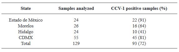
Conclusion. The new potexvirus was named Opuntia potexvirus A (OPV-A), whose viral replicase sequence has a 77.7 % amino acid identity with Schlumbergera virus X. Finally, CCV-1 was detected in 93 (72 %) of 129 vegetable prickly pear cactus samples collected in the four entities.

Keywords: Cactaceae, HTS, Viroid, Potexvirus, Tobamovirus, Carlavirus
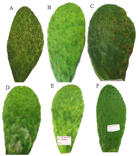
Figure 1. Symptoms associated with virosis in vegetable prickly pear cactus cladodes. (A-D) Samples of prickly pear cactus collected in Mexico City, Milpa Alta Borough, (E) Prickly pear cactus sample collected in the State of Mexico, Otumba Municipality, (F) Healthy prickly pear cactus sample from the State of Mexico.
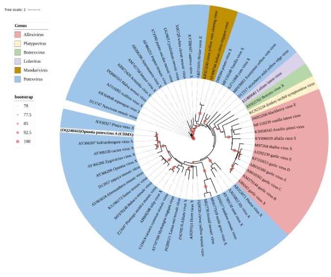
Figure 10. Maximum Likelihood phylogenetic tree of the Alphaflexiviridae family based on amino acid se- quences of the coat protein; the <em>Opuntia</em> potexvirus A sequence is highlighted in bold. Tree colors represent different genera within this family. Circles on branches indicate UFBoot support values >70%.
Figure 10. Maximum Likelihood phylogenetic tree of the Alphaflexiviridae family based on amino acid se- quences of the coat protein; the Opuntia potexvirus A sequence is highlighted in bold. Tree colors represent different genera within this family. Circles on branches indicate UFBoot support values >70%.
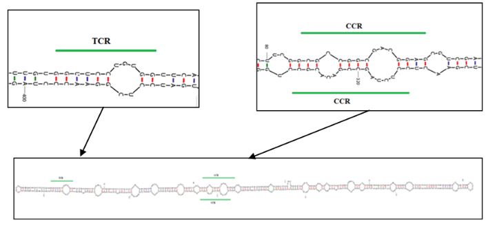
Figure 11. Predicted secondary structure using the UNAFold web server (UNAFold tool available online: www. mfold.org) for OVd-I. Highlighted are the sites forming the terminal conserved region (TCR) at positions 16 to 26 and the central conserved region (CCR) at positions 84 to 100 and 313 to 329
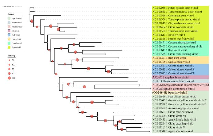
Figure 12. Maximum Likelihood phylogenetic tree of the Pospiviroidae and Avsunviroidae families based on nucleotide sequences of complete genomes from representative species of different genera. The sequence obtained from OVd-I is highlighted in bold. Various colors represent viroid genera. Circles on branches indicate UFBoot support values >50%.
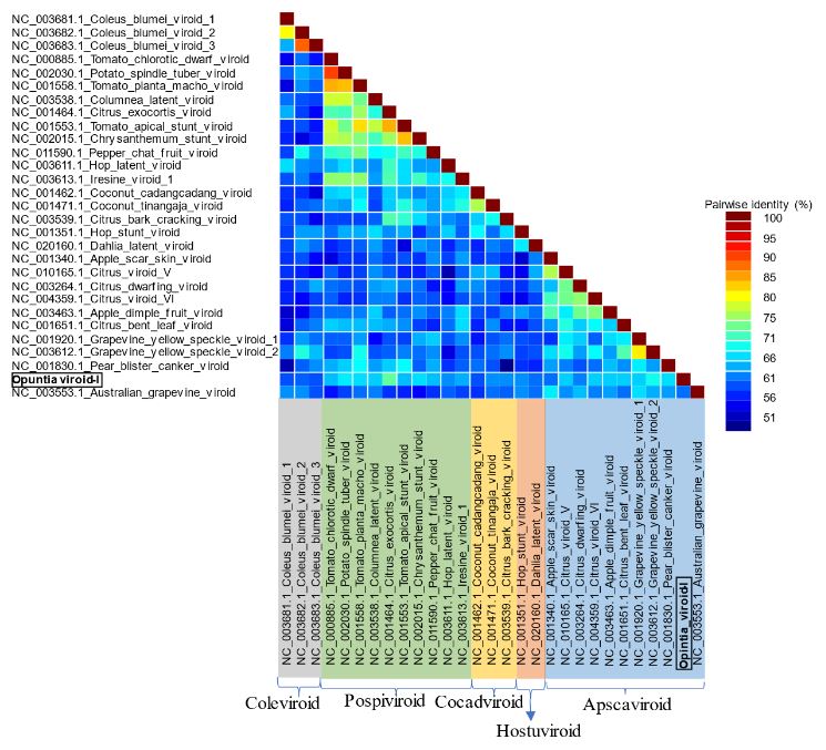
Figure 13. Pairwise identity frequency distribution obtained using the Sequence Demarcation Tool (SDT) (Muhire <em>et al.,</em> 2014) from complete genome sequences of Pospiviroidae family species to support the demarcation of <em>Opuntia</em> viroid I as a new species
Figure 13. Pairwise identity frequency distribution obtained using the Sequence Demarcation Tool (SDT) (Muhire et al., 2014) from complete genome sequences of Pospiviroidae family species to support the demarcation of Opuntia viroid I as a new species
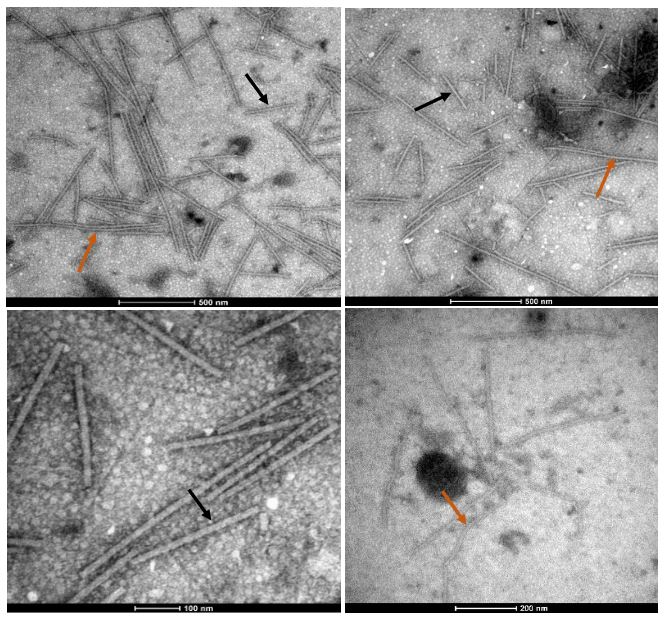
Figure 14. Viral particles in the form of rigid rods (®) and flexible filaments (®) of varying lengths were observed through transmission electron microscopy in vegetable prickly pear cactus exhibiting irregular yellow patterns, yellow rings, mosaic patterns, and chlorotic spots around the spines, employing the “leaf dip” technique
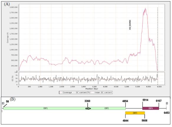
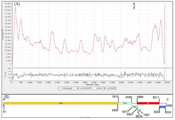
Figure 2. (A) Read coverage across the genome of the OV2-Edo-Mex isolate and the percentage of guanine-cytosine content, (B) Genomic map of OV2-Edo-Mex. The arrow indicates the termination codon site of the complete 128 kDa protein, with readthroughs
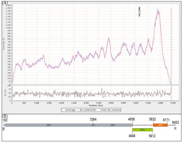
Figure 3. (A) Read coverage across the genome of the OV2-CDMX isolate and the percentage of guanine-cytosine content. (B) Genomic map of OV2-CDMX. The arrow indicates the termination codon site of the complete 128 kDa protein, with readthroughs.
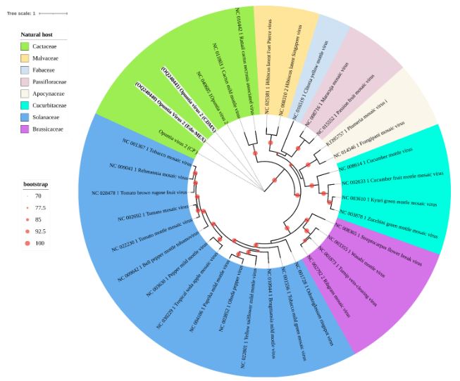
Figure 4. Maximum Likelihood phylogenetic tree of the Tobamovirus genus showing the relationships between genomes obtained in this study (in bold) for OV2 and those of previously reported isolates. Different colors represent botanical families as natural hosts where each virus has been reported. Circles on branches indicate UFBoot support values > 70%.
Figure 5. (A) Read coverage across the genome of the CCV-1-Edo-Mex isolate and the percentage of guanine-cytosine content. (B) Genome organization of CCV1-Edo-Mex, depicting six open reading frames and their corresponding products. RNA-dependent RNA polymerase (RdRp); coat protein (CP); nucleic acid-binding protein (NB); triple gene block (TGB)
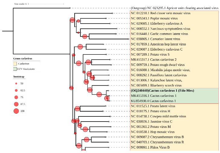
Figure 6. Maximum Likelihood phylogenetic tree of complete nucleotide sequences of CCV-1 genomes from this study (in bold), along with other isolates, and various species within the Carlavirus genus. Apricot vein clearing associated virus, Prunevirus genus, was used as an outgroup. Circles on branches indicate UFBoot support values > 50%.
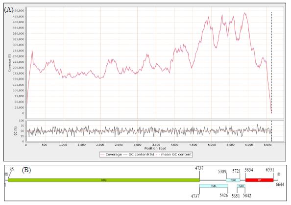
Figure 7. (A) Read coverage across the genome of <em>Opuntia potexvirus A (OPV-A)</em> and guanine/ cytosine percentage. (B) Genome organization of OPV-A, displaying five open reading frames and their corresponding products: RdRp, viral replicase; TGB, triple gene block; CP, coat protein.
Figure 7. (A) Read coverage across the genome of Opuntia potexvirus A (OPV-A) and guanine/ cytosine percentage. (B) Genome organization of OPV-A, displaying five open reading frames and their corresponding products: RdRp, viral replicase; TGB, triple gene block; CP, coat protein.
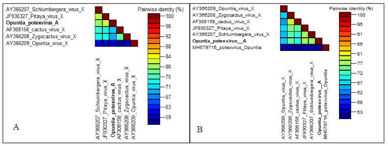
Figure 8. Matrix of amino acid identity percentage for the coat protein (A) and viral replicase (B) of species within the Potexvirus genus closest to <em>Opuntia</em> potexvirus A (in bold)
Figure 8. Matrix of amino acid identity percentage for the coat protein (A) and viral replicase (B) of species within the Potexvirus genus closest to Opuntia potexvirus A (in bold)
Figure 9. Maximum Likelihood phylogenetic tree of the Alphaflexiviridae family based on viral replicase amino acid sequences; the <em>Opuntia</em> potexvirus A sequence is highlighted in bold. Tree colors rep- resent different genera within this family. Circles on branches indicate UFBoot support values >70%
Figure 9. Maximum Likelihood phylogenetic tree of the Alphaflexiviridae family based on viral replicase amino acid sequences; the Opuntia potexvirus A sequence is highlighted in bold. Tree colors rep- resent different genera within this family. Circles on branches indicate UFBoot support values >70%
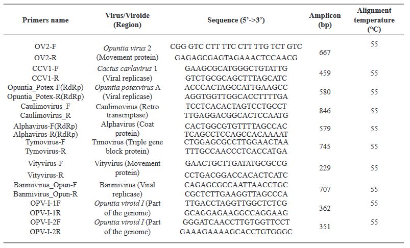
Table 1. Primers used in RT-PCR assays for the validation of viruses/viroids detected in the HTS data from two vegetable prickly pear cactus libraries
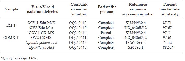
Table 2. Comparison of the complete or partial genome of the viruses identified through HTS in the two vegetable prickly pear cactus libraries with the most closely related reference sequence from GenBank.
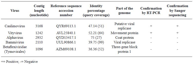
Table 3. Result of Blastx analysis for potentially present viruses in the CDMX-1 library, confirmed through RT-PCR and sequencing
Table 4. Detection of CCV-1 in four vegetable prickly pear cactus producing states in Mexico