RMF Logo

Article filters

Search Papers:

For searching before 2024 see Archive (2000-2023)
  • Vol. 2023, 41(4)
  • Phytopathological Note
  • Special Issue

Comparison of molecular protocols to detect Tomato brown rugose fruit virus in solanaceae hosts

By Erika J Zamora Macorra, Katia Aviña Padilla*, Rosemarie W Hammond, Daniel L Ochoa Martínez

*Corresponding Author.

Received: 23/August/2023 – Published: 23/December/2023
DOI: https://doi.org/10.18781/R.MEX.FIT.2023-5

  • PDF
  • HTML
  • XML

Abstract Background/Objective. The Tomato brown rugose fruit virus (ToBRFV) has emerged as a significant threat to Solanaceae crops, including tomato and pepper. Its presence in Mexico since 2018 has raised concerns about its impact on agricultural production. Early and accurate detection of this pathogen is essential to prevent its spread and mitigate its effects. In Mexico, several molecular techniques are employed for its diagnosis, including endpoint RT-PCR, RT-qPCR, and multiplex RT-qPCR.

Materials and Methods. This research aimed to assess the efficiency of different RNA extraction methods in combination with specific PCR primers for detecting ToBRFV.

Results. Among the methods tested, the CTAB-Trizol RNA extraction protocol combined with nested PCR using primers reported by Dovas et al. (2004) was identified as the most sensitive molecular method for detecting the virus.

Conclusion. This finding highlights the importance of selecting the appropriate combination of extraction and amplification protocols to achieve optimal sensitivity and accuracy in ToBRFV detection.

Keywords: Tomato brown rugose fruit virus, ToBRFV, Solanaceae crops, RNA extraction, RT-PCR, molecular detection, agricultural production
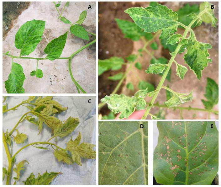
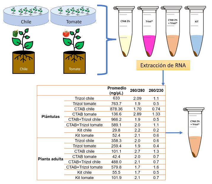
Figure 1. A-C) Symptoms of mosaic, leaf deformation, and leaf narrowing were evident in tomato saladette plants gathered from greenhouse settings. These plants tested positive for Tomato brown rugose fruit virus (ToBRFV); D-E) visual observations on inoculated Nicotiana leaves revealed the presence of localized chlorotic and necrotic lesions caused by ToBRFV infection. The virus-positive plants utilized for the study were sourced from Tecoman, Colima, Mexico.
Figure 2. The evaluation of total RNA extraction methods involved comparing their effectiveness and yield, as measured by absorbance readings on a Nanodrop 2000® spectrophotometer. Among the methods tested, including Trizol®, CTAB 2%, and CTAB 2%-Trizol®, the CTAB 2%-Trizol® protocol yielded the highest concentration of total RNA, while the RNA isolation Kit produced the lowest concentration. This data was recorded through absorbance measurements at wavelengths of 260/280 and 260/230. The Nanodrop 2000® was used to quantify the extracted RNA’s concentration and assess its quality by these absorbance ratios. The results indicated that the CTAB 2%-Trizol® protocol was particularly effective in extracting high-quality RNA from the plant samples, making it a suitable choice for subsequent molecular analyses.
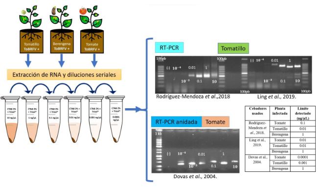
Figure 3. Evaluation and sensitivity of PCR primers. 1.5% agarose gels electrophoretic analysis of RT-PCR and Nested RT-PCR products (Ling´s primers expected size 842 pb; Rodríguez-Mendoza´s primers expected size 475 bp and Dovas´s primers expected size 400 pb). (-): sterilized water instead RNA. 100 bp = 100bp DNA Ladder (Invitrogen®). 1Kb= 1000 bp DNA ladder (Promega®). 10-3 and 10-4 = 0.001 and 0.0001 ng μL-1.
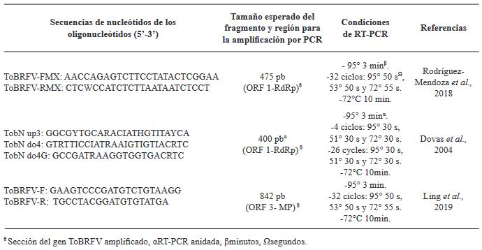
Table 1. Primers tested in this study for ToBRFV detection in tomato, tomatillo, and eggplant
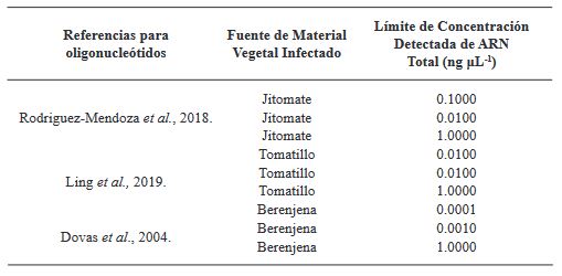
Table 2. Comparison of Primer References, Plant Sources, and RNA Detection Limits in source of infected plant material