Genus Orthotospovirus in Costa Rica: A Central American case
By
Mauricio Montero Astúa*,
Natasha Dejuk Protti,
David Bermúdez Gómez,
Elena Vásquez Céspedes,
Laura Garita Salazar,
Federico J Albertazzi,
Scott Adkins ,
Lisela Moreira Carmona
*Corresponding Author.
Received: 23/August/2023 – Published:
28/December/2023
DOI:
https://doi.org/10.18781/R.MEX.FIT.2023-6
Abstract Background/Objective. The Orthotospovirus genus encompasses a range of economically significant and emerging plant viruses that affect a variety of crops globally. While the prevalence and characteristics of these phytopathogenic viruses are extensively documented in North and South America, their presence in Central America remains comparatively underexplored. This study focuses on Costa Rica, strategically positioned at the nexus of North and South America, to enhance our understanding of Orthotospovirus in this region.
Materials and Methods. We analyzed 295 plant samples using enzyme-linked immunosorbent assay (ELISA) to test for the presence of INSV, IYSV, TSWV, and the GRSV/TCSV serogroup. Additionally, a subset (20 samples) underwent further scrutiny through reverse transcription-polymerase chain reaction (RT-PCR) employing both universal and species-specific primers.
Results. Our ELISA results indicated the absence of TSWV and the GRSV/TCSV serogroup. However, the presence of INSV in Costa Rica was substantiated through ELISA, RT-PCR, and partial sequencing, revealing its prevalence in both open-field and greenhouse environments. Despite previous diagnostic reports suggesting the presence of TSWV in Costa Rica, our study did not detect this virus. RT-PCR analysis with degenerate primers also found no evidence of other Orthotospovirus species in our samples. The identification of a dominant INSV haplotype, along with three additional variants, suggests the likelihood of at least two independent virus introductions into the region.
Conclusion. These findings underscore the necessity for more comprehensive surveys and research on Orthotospoviruses in Central America to better understand their epidemiology and impact on agriculture.
Keywords:
viral symptoms, ELISA, RT-PCR, genetic diversity, INSV, IYSV
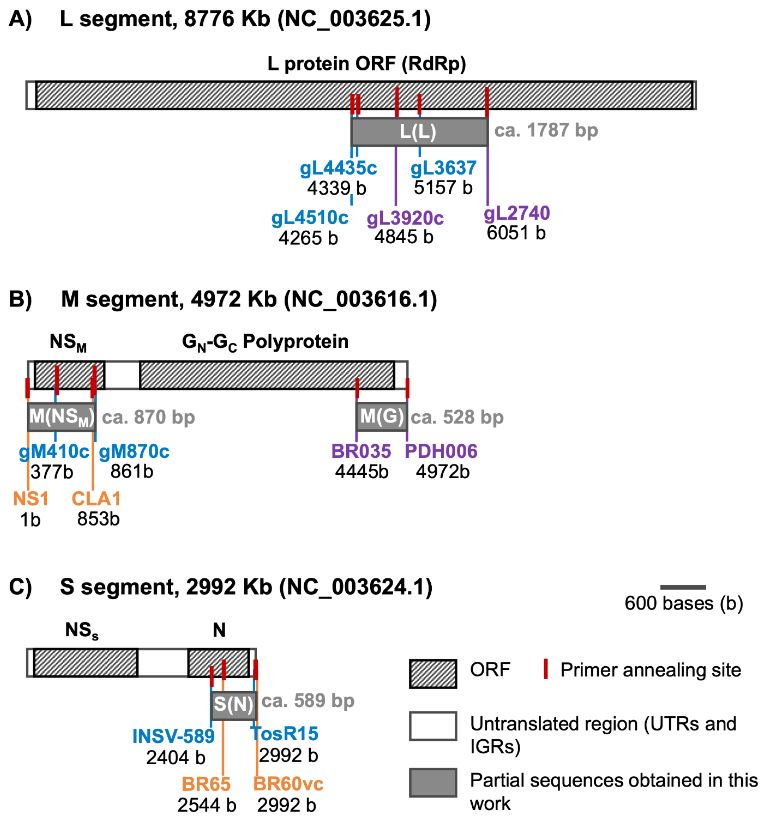
Figure 1. Representation of primer pairs annealing positions in reference to the complete genome of Impatiens necrotic spot virus; accession numbers in parenthesis for each genome segment. Overlapping among primers and the final partial sequences (gray rectangles) obtained herein with corresponding expected size (bp). Base pair values under each primer represent the positions of the first and last nucleotide of the corresponding amplicon in the reference genome

Figure 2. Diverse Symptoms of Impatiens necrotic spot virus (INSV) in Multiple Hosts Confirmed by ELISA and RT-PCR/Sequencing. A: Necrotic concentric rings on Impatiens (Impatiens sp.) T031. B, C: Chlorotic rings and spots on Snapdragons (Antirrhinum majus) T057. D: Chlorotic and necrotic striate on orchid leaves T024. E, F: Chlorotic mosaic on Amaryllis (Hippeastrum sp.) T161 and Iris (Iris sp.) T184. G: Necrotic concentric rings on Coleus (Plectranthus scuttellarioides) T157. H: Chlorotic patterns on Basil (Ocimum basilicum) T156. I, M: Chlorotic concentric ring patterns on Sweet Pepper (Capsicum annuum) leaves T250 and T092. J, K, L: Uneven ripening, ring patterns, and deformation on Sweet Pepper fruits T092, T107, T094. N, O: Chlorotic patterns, rings, and uneven surface on Tomato (Solanum lycopersicum) fruits T263 and T303.

Figure 3. Long-Term Detection of impatiens necrotic spot and tomato spotted wilt viruses by ELISA. This graph presents the frequency of detection for both Impatiens necrotic spot virus and Tomato spotted wilt virus over a 23.5-year period (2000 to July 2023). The data, derived from samples submitted to the diagnostic services at the Cellular and Molecular Biology Research Center, University of Costa Rica, illustrate the temporal trends and occurrence patterns of these viruses in Costa Rica.
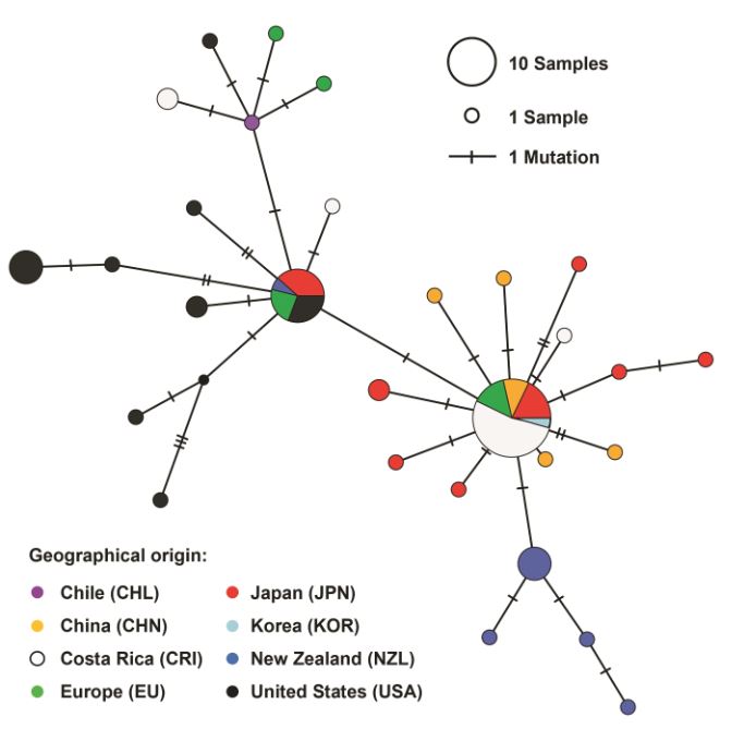
Figure 4. Haplotype Network Graph of Impatiens Necrotic Spot Virus. This graph is a haplotype network inferred using the TCS method, based on a 261 bp sequence alignment of the nucleocapsid protein ORF (S[N] segment) of Impatiens necrotic spot virus. The network includes data from 81 isolates, visually representing the genetic relationships and diversity among the different haplotypes of the virus.
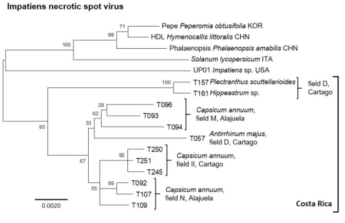
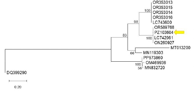
Figure 5. Comprehensive phylogenetic analysis of impatiens necrotic spot virus isolates. This figure illustrates a phylogenetic tree constructed from an alignment of concatenated sequences (2945 positions) encompassing regions of the nucleocapsid [S(N)], glycoprotein precursor [M(G)], movement protein [M(NSM)], and viral polymerase [L(L)] ORFs. The tree includes isolates from various countries, identified by ISO 3166-1 three- letter codes: CHN (China), ITA (Italy), KOR (South Korea), and USA (United States of America). For Costa Rican isolates, additional distinct letter codes in parenthesis signify separate geographic locations. The analysis, executed in MEGA X, utilized the Maximum Likelihood method with a Tamura-3-parameter model and a gamma-distributed rate of variation in nucleotides (+G), involving 2000 permutations. The scale bar indicates the number of nucleotide substitutions per site.

Table 4. Partial nucleotide sequences and virus identity determined for a subset of INSV ELISA-positive samples
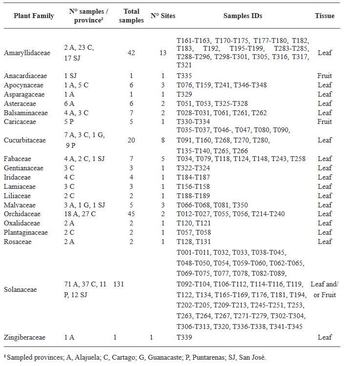
Table 1. Samples collected from five provinces of Costa Rica to analyze the presence of species of Orthotospvirus
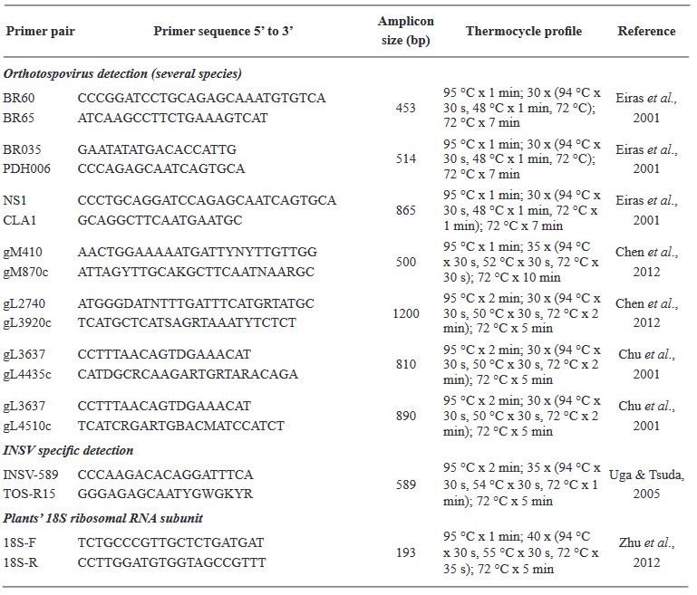
Table 2. Primer pairs and thermocycle profiles used for the detection of Orthotospoviruses
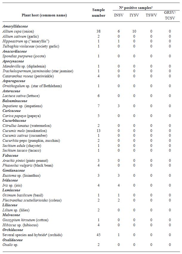
Table 3. Number of total and positive samples per plant species tested by ELISA to detect three Orthotospoviruses in Costa Rica.