RMF Logo

Article filters

Search Papers:

For searching before 2024 see Archive (2000-2023)
  • Vol. 2025, 43(2)
  • Scientific Article

Cloning, expression, and serological detection of the putative RNA polymerase of Papaya meleira virus Mexican variant in Escherichia coli

By Wendy Serra, Jeanin A Arguelles Quintal, Mildred Carrillo Pech, Alethia F Toriz , Enrique Castaño, Ignacio Islas Flores, Luisa A Lopez Ochoa*

*Corresponding Author.

Received: 01/May/2024 – Published: 30/April/2025
DOI: https://doi.org/10.18781/R.MEX.FIT.2405-00

  • PDF
  • HTML
  • XML

Abstract Background/Objective. Papaya meleira disease in Mexico is associated to Papaya meleira virus Mexican variant (PMeV-Mx) and is characterized by the spontaneous exudation of latex in fruits. PMeV-Mx ORF2 encodes a protein with RNA-dependent RNA polymerase (RdRp) motifs, which is essential for viral replication. This study aimed to develop a method for producing and purifying recombinant PMeV-Mx ORF2 encoded protein (pORF2) in E. coli and generate specific antibodies for its detection.

Materials and Methods. The PMeV-Mx genome was analyzed using UGENE to predict ORFs and identify the putative slippery site. ORF1 and ORF2 were amplified by PCR from cDNA obtained from infected papaya latex, cloned into pGEM-T Easy, and subsequently transferred into pDONR221 and pDEST17 for expression in E. coli. Recombinant 6xHis-pORF2 was expressed in E. coli BL21 strain, induced with IPTG, and purified under denaturing conditions. The purified protein was used to generate polyclonal antibodies, with immunizations conducted at different time points. Antisera specificity and optimal working dilutions were evaluated by immunodetection assays, using recombinant 6xHis-pORF2 as the target and His-tagged proteins as negative controls. Additionally, papaya plants were inoculated with latex from symptomatic fruits as virus reservoir and PMeV-Mx infection was confirmed by RT-PCR.

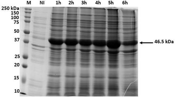
Results. The recombinant 6xHis-pORF2 protein of PMeV-Mx was expressed in E. coli and purified. A ~46.5 kDa band was detected, consistent with its estimated molecular weight. The protein expression increased between 2- and 6-hours post-induction with IPTG. Western blot analysis confirmed the presence of the His tag and the integrity of the recombinant protein. Purification using Ni-NTA resin resulted in a strong ~46.5 kDa band along with light bands ranging from 15 to 150 kDa. Polyclonal antibodies against pORF2 were generated and specifically recognized the purified protein in immunoassays, with detection observed at dilutions from 1:500 to 1:3000. No cross-reactivity was observed with negative controls, but a non-specific ~75 kDa band was detected in E. coli extracts.

Conclusion. A protocol for expressing, detecting, and purifying the recombinant 6xHis-pORF2 protein from PMeV-Mx in E. coli was established. Rabbit polyclonal antibodies recognized the target protein in bacterial fractions, though further optimization is needed to enhance specificity. These antibodies will support future diagnostic development and protein characterization in plants.

Keywords: Papaya meleira, Recombinant proteins, Antibodies, Umbra-like viruses, PMeV-Mx
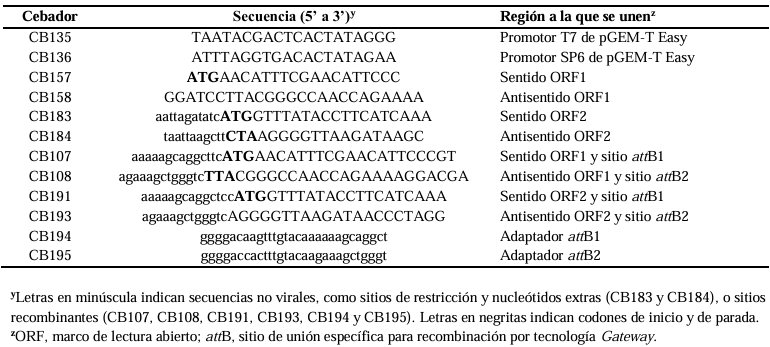
Table 1. Primers used TO clone PMeV-Mx ORF1 AND ORF2 IN pDEST17 vector, TO generate plasmids pCB252 AND pCB253, respectively.
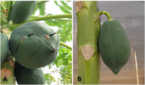
Figure 1. Fruits of <em>Carica papaya</em> cv. Maradol plants grown in the screen house. A) Papaya fruits infected with PMeV-Mx showing the typical meleira disease symptom of spontaneous latex exudation. B) Fruit of mock-inoculated papayas (Healthy).
Figure 1. Fruits of Carica papaya cv. Maradol plants grown in the screen house. A) Papaya fruits infected with PMeV-Mx showing the typical meleira disease symptom of spontaneous latex exudation. B) Fruit of mock-inoculated papayas (Healthy).
Figure 2. Genomic organization of PMeV-Mx. Numbers indicate the nucleotide (nt) positions and the total number of nt in the genome. Solid boxes in the genome represent ORF1 and ORF2, respectively. Solid boxes below indicate the putative proteins, product of ORF1 and ORF2 (pORF1 and pORF2, respectively) and the putative fusion product of -1 ribosomal frameshifting (pORF1/pORF2). The arrow indicates the position of the heptanucleotide.
Figure 3. Time course of PMeV-Mx 6xHis-pORF2 expression using insoluble crude <em>E. coli</em> lysate to establish the optimal induction period for expression. Expression of 6xHis-pORF2 was induced with 1 mM IPTG, at 37 ºC. Aliquots were removed at the times indicated (1 to 6 h after IPTG induction) and proteins were visualized by Coomassie-stained SDS gel. M, Bio-Rad 10-250 kDa marker; NI, non-induced control. The arrow indicates the 46.5 kDa 6xHis-pORF2 protein.
Figure 3. Time course of PMeV-Mx 6xHis-pORF2 expression using insoluble crude E. coli lysate to establish the optimal induction period for expression. Expression of 6xHis-pORF2 was induced with 1 mM IPTG, at 37 ºC. Aliquots were removed at the times indicated (1 to 6 h after IPTG induction) and proteins were visualized by Coomassie-stained SDS gel. M, Bio-Rad 10-250 kDa marker; NI, non-induced control. The arrow indicates the 46.5 kDa 6xHis-pORF2 protein.
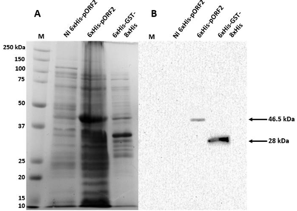
Figure 4. Detection of PMeV-Mx 6xHis-pORF2 in <em>E. coli</em> crude lysates by monoclonal anti-His antibodies. A) Coomassie stained SDS gel. B) Western blot in PVDF membranes incubated with a 1:1000 dilution of the anti-His tag monoclonal antibody coupled to horseradish peroxidase (HRP). M, Bio-Rad 10-250 kDa marker; NI and 6xHis GST-8xHis, non-induced and positive controls, respectively; 6xHis-pORF2, cellular fraction with recombinant PMeV-Mx pORF2 expressed in BL21 cells. The arrows indicate the 46.5 kDa 6xHis-pORF2 and the 28 kDa 6xHis GST-8xHis proteins, respectively.
Figure 4. Detection of PMeV-Mx 6xHis-pORF2 in E. coli crude lysates by monoclonal anti-His antibodies. A) Coomassie stained SDS gel. B) Western blot in PVDF membranes incubated with a 1:1000 dilution of the anti-His tag monoclonal antibody coupled to horseradish peroxidase (HRP). M, Bio-Rad 10-250 kDa marker; NI and 6xHis GST-8xHis, non-induced and positive controls, respectively; 6xHis-pORF2, cellular fraction with recombinant PMeV-Mx pORF2 expressed in BL21 cells. The arrows indicate the 46.5 kDa 6xHis-pORF2 and the 28 kDa 6xHis GST-8xHis proteins, respectively.
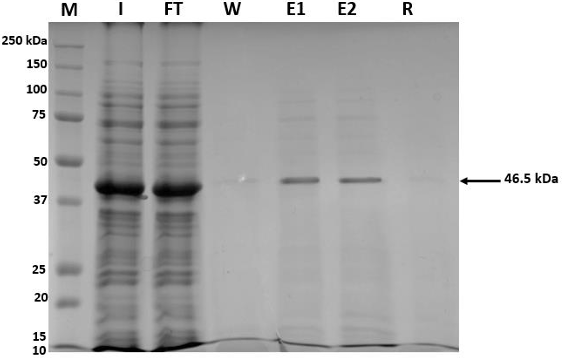
Figure 5. Purification of PMeV-Mx 6xHis-pORF2 with Ni-NTA Agarose under denaturing conditions. M, Bio-Rad 10-250 kDa marker; I, <em>input</em>; Ft, <em>flow-through</em>; E1 and E2, first and second eluates, respectively; R, resin. The arrow indicates the 46.5 kDa 6xHis-pORF2 protein.
Figure 5. Purification of PMeV-Mx 6xHis-pORF2 with Ni-NTA Agarose under denaturing conditions. M, Bio-Rad 10-250 kDa marker; I, input; Ft, flow-through; E1 and E2, first and second eluates, respectively; R, resin. The arrow indicates the 46.5 kDa 6xHis-pORF2 protein.
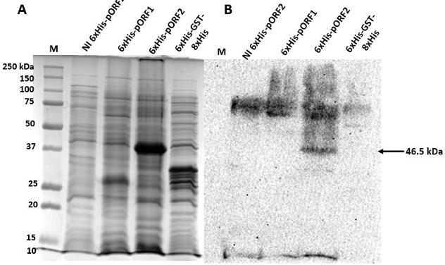
Figure 6. Detection of PMeV-Mx 6xHis-pORF2 in <em>E. coli</em> crude lysate with anti-pORF2 antibodies. A) Coomassie-stained SDS gel. B) Western blot (Nitrocellulose membrane) incubated with the anti- PMeV-Mx pORF2 polyclonal antibody (dilution 1:1000), and the anti-rabbit coupled to horseradish peroxidase (HRP) secondary antibody (dilution 1:3000). M, Bio-Rad 10-250 kDa marker; NI, non-induced cellular fraction. 6xHis-pORF1 and 6xHis GST-8xHis were used as negative controls. The arrow indicates the 46.5 kDa 6xHis-ORF2 protein.
Figure 6. Detection of PMeV-Mx 6xHis-pORF2 in E. coli crude lysate with anti-pORF2 antibodies. A) Coomassie-stained SDS gel. B) Western blot (Nitrocellulose membrane) incubated with the anti- PMeV-Mx pORF2 polyclonal antibody (dilution 1:1000), and the anti-rabbit coupled to horseradish peroxidase (HRP) secondary antibody (dilution 1:3000). M, Bio-Rad 10-250 kDa marker; NI, non-induced cellular fraction. 6xHis-pORF1 and 6xHis GST-8xHis were used as negative controls. The arrow indicates the 46.5 kDa 6xHis-ORF2 protein.