RMF Logo

Article filters

Search Papers:

For searching before 2024 see Archive (2000-2023)
  • Vol. 2025, 43(1)
  • Phytopathological Note

Etiology of brown rot in strawberry (Fragaria x nanassa) in the State of Mexico

By Hugo Velasco Montaño, Victoria Ayala Escobar, Daniel Téliz Ortiz, Nadia Landero Valenzuela*, Santos Gerardo Leyva Mir

*Corresponding Author.

Received: 07/June/2024 – Published: 27/December/2024
DOI: https://doi.org/10.18781/R.MEX.FIT.2406-3

  • PDF
  • HTML
  • XML

Abstract Background/Objective. In a strawberry crop established in a greenhouse in Montecillo, Texcoco, State of Mexico, in 2022, brownish brown leaf spots and rotting of fruits with asymmetric sunken lesions were observed, which extended and acquired a brown color. The objective of the present work was to identify the causal agent of brown rot in fruits and strawberry plants.

Materials and Methods. Symptomatic fruits and leaves were collected, from which fungal isolates were obtained to perform pathogenicity tests on plants and fruits, in plants by two inoculation methods: spraying via foliar and via root; in fruits by immersion. Concentrations of 2×10⁶ conidia mL-1 were used. The ITS region of the rDNA was amplified and sequenced by PCR with the universal primers ITS1-ITS4.

Results. Pilidium concavum was morphologically and molecularly identified as the causal agent of brown spot and brown rot on strawberry. It was found to be pathogenic in strawberry fruits cv. Aromas and in plants less than two months old. It showed variation in virulence, in affected plants it varied from 40 to 50%, in fruits it reached 100%.

Conclusion. The result determines that Pilidium concavum is a pathogen that produces brown leaf spot and brown rot in strawberry fruits. It allows new lines of research related to the impact of the disease on strawberry production, yield and quality in Mexico. This research is the first report of Pilidium concavum as a strawberry pathogen in the State of Mexico.

Keywords: Pilidium concavum, strawberry, pathogenicity, virulence
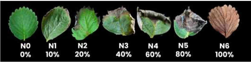
Figure 1. Visual scale for evaluating the severity of brown toasted leaf spot on strawberry. Designed by the authors.
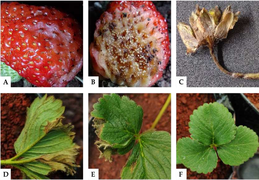
Figure 2. A) Fruit with sunken, wet lesions. B) Fruit with brown rot and fungal structures. C) Brown constriction on the flower peduncle. D-E) Leaves with toasted brown edges. F) Healthy leaves.
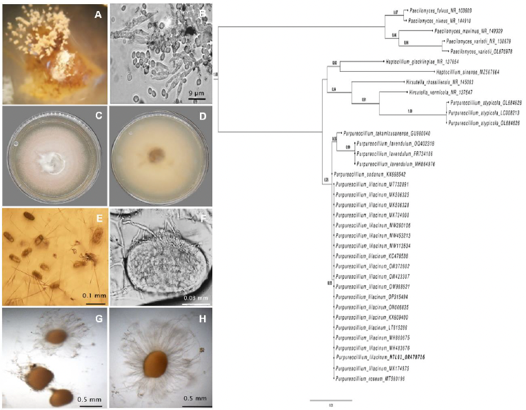
Figure 3. A) Sporodochia on strawberry fruit. B) Sporodochia on the underside of the leaf. C-D) Gelatinous colony with conidial mass on PDA medium. E-F) Longitudinal and transverse sections of the sporodochium. G) Hyaline, cylindrical, filiform conidiophores. H) Hyaline, aseptate, allantoid conidia.
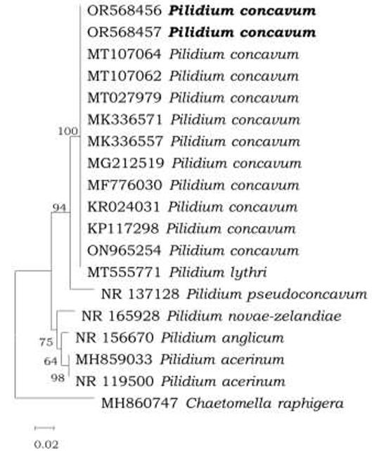
Figure 4. Phylogenetic tree based on Neighbor Joining of the ITS rDNA sequence, showing a phylogenetic affinity of the Mexico isolate (in bold) with <em>Pilidium concavum</em> above 95% of the node. Bar scaling 0.02 represents nucleotide substitutions per site.
Figure 4. Phylogenetic tree based on Neighbor Joining of the ITS rDNA sequence, showing a phylogenetic affinity of the Mexico isolate (in bold) with Pilidium concavum above 95% of the node. Bar scaling 0.02 represents nucleotide substitutions per site.
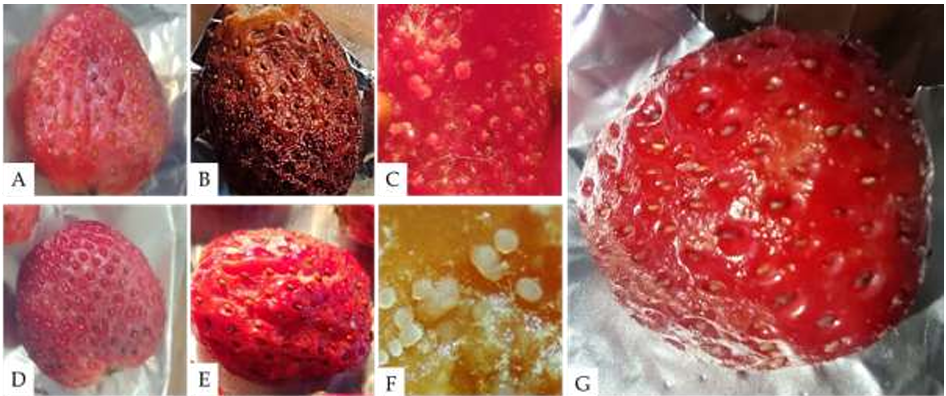
Figure 5. Pathogenicity tests on strawberry fruit inoculated with a concentration of 2×10<sup>6</sup> conidia mL<sup>−1</sup> of <em>Pilidium concavum</em>. A) Fruit with a wound inoculated with the fungus. B) Wounded fruit showing symptoms 72 hours post-inoculation (hpi). C) Sporodochia of the fungus on the wounded fruit. D) Unwounded fruit inoculated with the fungus. E) Unwounded fruit showing symptoms 96 hours post-inoculation. F) Sporodochia of the fungus on the unwounded fruit. G) Control inoculated with sterile distilled water, showing no symptoms at 96 hours post-inoculation.
Figure 5. Pathogenicity tests on strawberry fruit inoculated with a concentration of 2×10⁶ conidia mL−1 of Pilidium concavum. A) Fruit with a wound inoculated with the fungus. B) Wounded fruit showing symptoms 72 hours post-inoculation (hpi). C) Sporodochia of the fungus on the wounded fruit. D) Unwounded fruit inoculated with the fungus. E) Unwounded fruit showing symptoms 96 hours post-inoculation. F) Sporodochia of the fungus on the unwounded fruit. G) Control inoculated with sterile distilled water, showing no symptoms at 96 hours post-inoculation.
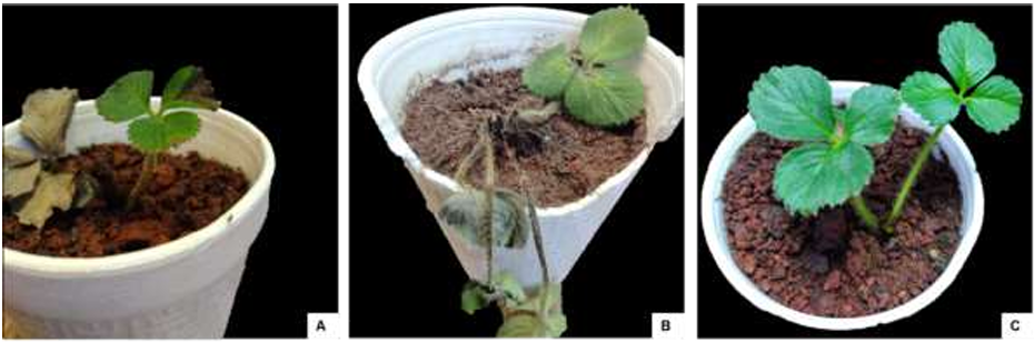
Figure 6. Strawberry plants (<em>Fragaria</em> x <em>ananassa</em>) cv. Aromas less than three months old, 15 days post-inoculation (dpi) with a suspension of 2x10<sup>6</sup> conidia per mL of <em>Pilidium concavum</em>. A) Inoculated via foliar; B) Inoculated via root routes; C) Control inoculated with sterile distilled water. 
Figure 6. Strawberry plants (Fragaria x ananassa) cv. Aromas less than three months old, 15 days post-inoculation (dpi) with a suspension of 2x10⁶ conidia per mL of Pilidium concavum. A) Inoculated via foliar; B) Inoculated via root routes; C) Control inoculated with sterile distilled water. 
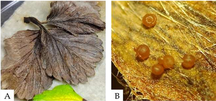
Figure 7. A) Asymptomatic strawberry leaves after 28 days post-inoculation (dpi), placed in a humid chamber. B) Formation of sporodochia on the leaves after 5 days in the humid chamber.