Share this link via
Or copy link




Article filters
Search Papers:
byElena Lugo García, Alejandro Salinas Castro, Gandhi Landa Cadena, Claudia Martínez Anaya, Irán Tapia Vázquez*
Received: 07/May/2026 – Published: 25/August/2026
DOI: https://doi.org/10.18781/R.MEX.FIT.2505-3
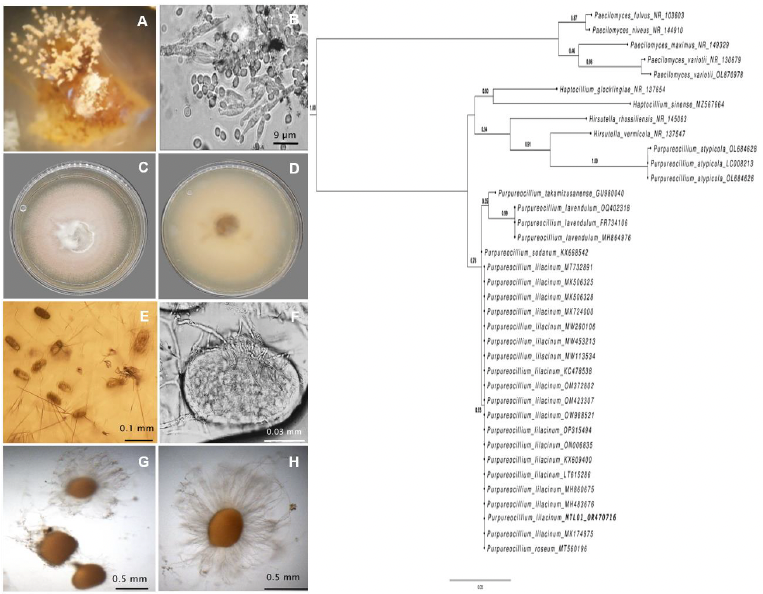
Abstract Background/Objective. The aim of this study was to determine the combined effect of an environmental strain of Purpureocillium lilacinum (MTL01) together with Bacillus velezensis 83, upon infection by the root-knot nematode Meloidogyne enterolobii.
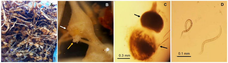
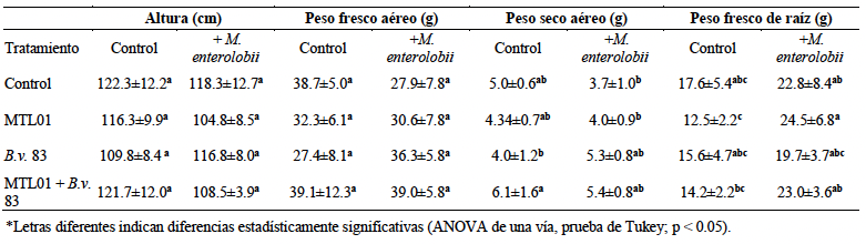
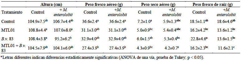
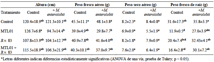
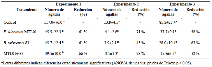
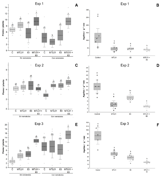
Experimental development. Three greenhouse experiments were conducted at different times (July–October 2022, February–June 2023, and September 2024–January 2025). In each experiment, treatments included uninoculated controls, plants inoculated only with P. lilacinum MTL01, with B. velezensis 83 only, or with both microorganisms, with and without nematode infection. Certified tomato seeds were inoculated with MTL01 spores or solution as a control and transplanted after germination. B. velezensis 83 was inoculated one week later, followed by two additional applications. Nematodes were introduced three weeks after transplanting, and plants were subsequently maintained in a greenhouse for an additional eight weeks. At harvest, growth parameters and infection levels were quantified (biomass, length, number of flowers, root weight, number of galls, and galling index).
Results. Data of vegetative growth varied among experiments, with no clear evidence of effects associated with microbial inoculation. However, the combination of P. lilacinum MTL01 and B. velezensis 83 showed reproducible results, reducing gall formation caused by M. enterolobii by more than 60% and promoting a consistent tendency toward increased flowering relative to nematode-infested control plants. These results demonstrate the potential of using combined microorganisms to improve tomato performance under greenhouse conditions.
Conclusion. Our findings highlight the importance of mitigating M. enterolobii infection in tomato using a combination of microorganisms, not only to control nematode populations but also to increase flower yield by up to 4.3-fold (Experiment 3) in plants without nematodes, and 1.6-fold in the presence of M. enterolobii. This represents a promising strategy; however, further research is needed to confirm its safety and optimize its application under real agricultural conditions.
Share this link via
Or copy link
byMiguel José Luna Alvarez, Martin Aguilar Tlatelpa, Marja Liza Fajardo Franco*
Received: 13/March/2026 – Published: 21/August/2026
DOI: https://doi.org/10.18781/R.MEX.FIT.2603-3
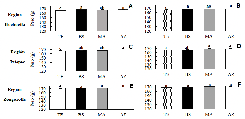
Abstract Background/Objective. Coffee leaf rust is the primary disease affecting this crop. The objective was to evaluate the incidence and severity of coffee leaf rust under the influence of a systemic fungicide and two biofungicides on varieties cultivated in the Northern and Northeastern Sierra regions of Puebla.
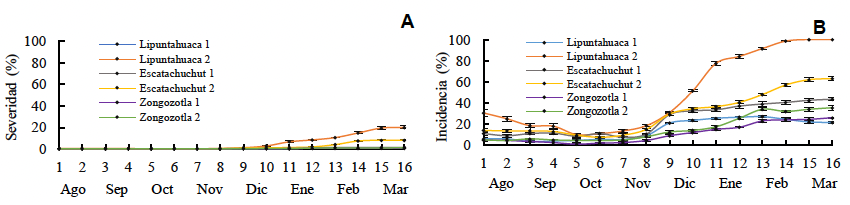
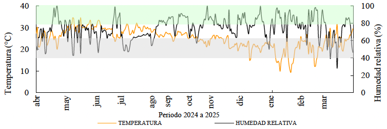
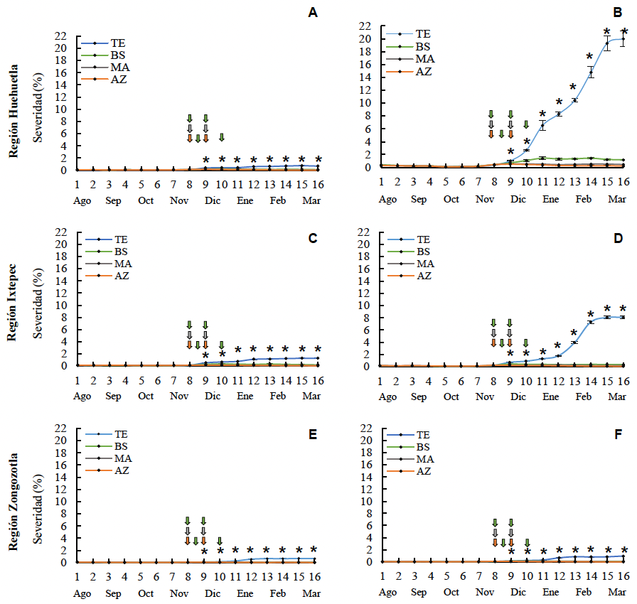
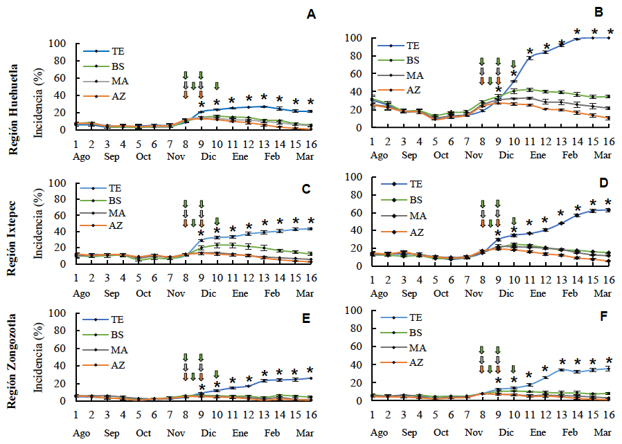
Experimental development. The effects of Azoxystrobin, Melaleuca alternifolia, and Bacillus subtilis were evaluated across six shade-grown plots featuring the Typica, Costa Rica 95, Garnica, Marsellesa, and Oro Azteca varieties, located in Huehuetla, Ixtepec, and Zongozotla. Sixteen sampling events were conducted, for each, of evaluation rust incidence and severity, shade percentage, the weight of 100 ripe cherries, and altitude were assessed. Additionally, temperature and relative humidity were recorded in Huehuetla. Incidence and severity were compared using the Kruskal-Wallis test and the Dwass-Steel-Critchlow-Fligner (DSCF) method. Furthermore, the correlation of these variables with altitude and shade percentage was analyzed. Cherry weight data underwent analysis of variance and mean comparison, while a risk analysis was performed using the climatic variables.
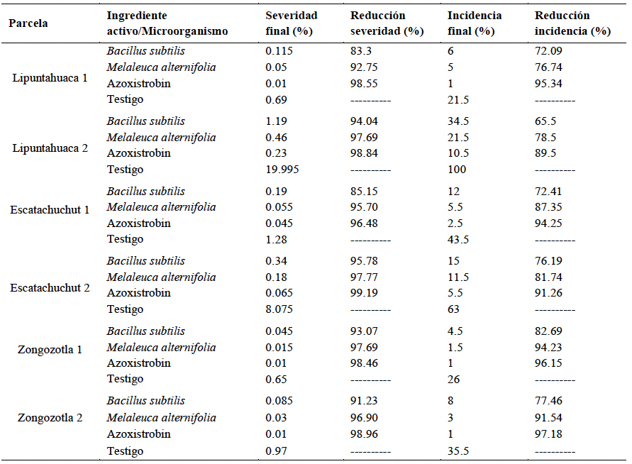
Results. Rust reached maximum severity and incidence (19.9 and 100%, respectively) in the Typica variety (March 2025). Azoxystrobin significantly reduced the disease, followed by M. alternifolia and B. subtilis. No influence of shade on incidence (r=0.33360) or severity (r=0.30735) was identified. In contrast, altitude showed a moderate negative correlation with incidence (r=-0.4632, p<0.0001) and with severity (r=-0.57433, p<0.0001). The Typica and Garnica varieties were the most susceptible.
Conclusion. The fungicides demonstrated control of the disease compared to the control treatment; therefore, they could be considered for the management of H. vastatrix.
Share this link via
Or copy link

First detection of bell pepper endornavirus infecting bell pepper in Guanajuato, Mexico
byAna Belen Duarte Cruz, Víctor Manuel Martín del Campo Soler, Kristian Stevens, Alfredo Diaz Lara*
Received: 25/May/2026 – Published: 21/August/2026
DOI: https://doi.org/10.18781/R.MEX.FIT.2605-3
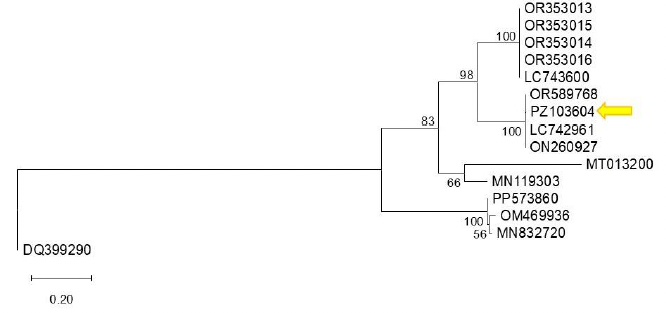
Abstract Background/Objective. Bell pepper (Capsicum annuum) is one of the most important agricultural crops of Mexico. Globally, over 60 virus species have been reported infecting Capsicum spp., some of them are associated with significant losses in yield and quality. The objective of this report is to determine the presence of bell pepper endornavirus (BPEV, Alphaendornavirus capsici) in bell pepper crops in Guanajuato, Mexico.
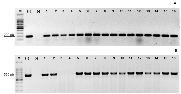
Experimental development. Samples of bell pepper plants were collected from a commercial greenhouse in Guanajuato, Mexico, in May 2024. A symptomatic plant (mosaic, leaf yellowing, and dwarfing symptoms) was analyzed by high throughput sequencing (HTS). Detection and identification were performed on individual samples using reverse transcription PCR (RT-PCR) and corroborated by Sanger sequencing. A phylogenetic analysis was conducted to investigate genetic diversity of the virus.
Results. A partial sequence of BPEV was generated by HTS and de novo assembly (GenBank: PZ103604), this virus was part of a mixed infection in the symptomatic plant, including Begomovirus capsicumhuastecoense (pepper huasteco yellow vein virus) and Cucumber mosaic virus. In addition, five bell pepper plants, including symptomatic and asymptomatic, tested positive for BPEV using RT-PCR, the identity of the virus was confirmed through Sanger sequencing and phylogenetic analysis.
Conclusion. The detection of BPEV in commercial bell pepper crops in Guanajuato highlights the importance of continuous monitoring of emerging viruses, including possible mixed infections with other viruses, and further studying their impact on crop health and productivity. Although BPEV was detected in symptomatic plants, the observed symptoms cannot be attributed to it as it was detected as part of a co-infection. To our knowledge, this is the first report of BPEV infecting bell pepper in Mexico.
Share this link via
Or copy link
byJosé Alan Reyes Pérez, Victorino Morales Ramos*, Ma. de Lourdes Catalina Arévalo Galarza, Dora Angélica Avalos de la Cruz
Received: 09/February/2026 – Published: 13/August/2026
DOI: https://doi.org/10.18781/R.MEX.FIT.2602-2
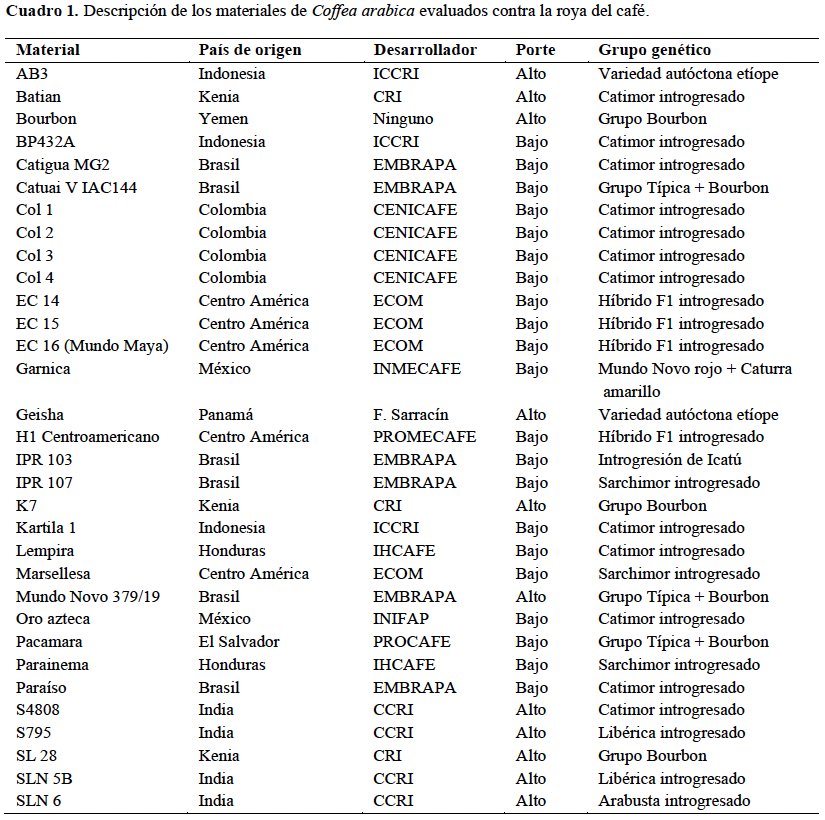
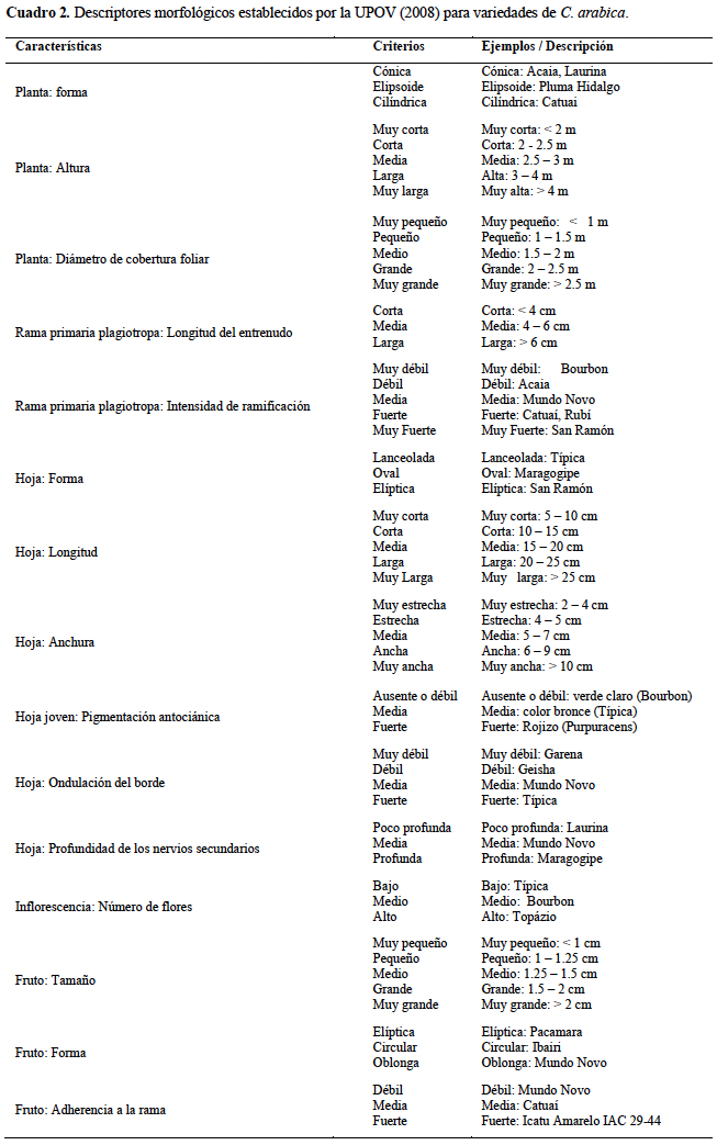
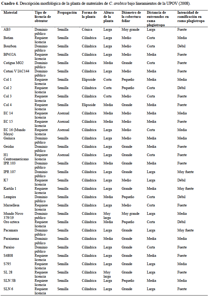
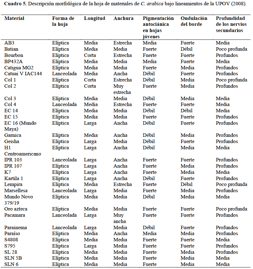
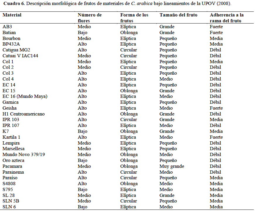
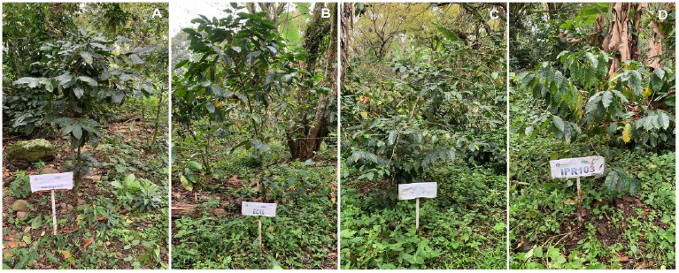
Abstract Backgrounds/Objective. Coffee cultivation is at risk due to the effects of climate change, which has created favorable conditions for the proliferation of diseases such as coffee leaf rust (Hemileia vastatrix). One of the most cost-effective and efficient control measures has been the use of materials that are tolerant or resistant to this fungus. The objective of this study was to analyze the response of 32 promising Coffea arabica accessions to coffee leaf rust and to describe their morphological characteristics, relating them to disease severity, under conditions prevailing in the municipality of Amatlán de los Reyes, Veracruz.
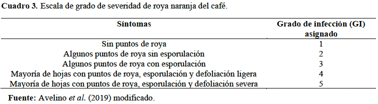
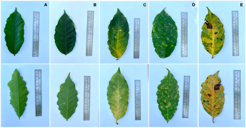
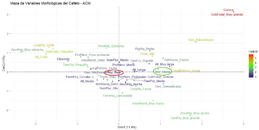
Experimental development. A total of 32 C. arabica accessions were evaluated in a completely randomized block design with three replications, compiling a descriptive profile of the visible, identifiable, and measurable morphological traits of each evaluated accession. The incidence of coffee leaf rust (Hemileia vastatrix) was assessed on a five-level severity scale on a monthly basis over a two-year period. An ordinal mixed-effects model and post hoc tests were computed to identify significant differences in rust incidence. Hierarchical cluster analysis was carried out to group the materials according to susceptibility or severity level, and the relationship between morphological traits and infestation level was analyzed using multiple correspondence analysis.
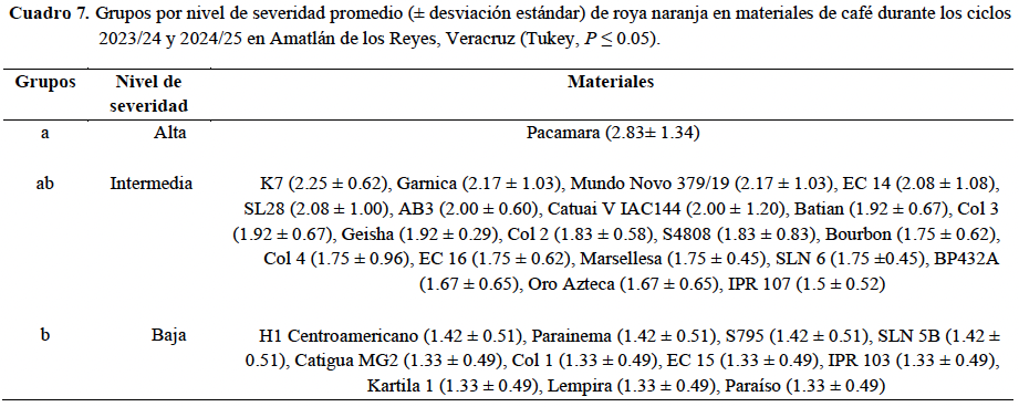
Results. The materials showed significant differences in tolerance to coffee leaf rust. Tukey's test (α = 0.05) identified three groups based on the severity of rust damage; the Pacamara variety was the most affected, while the H1 Centroamericano, Parainema, Catigua MG2, Col 1, EC 15, IPR 103, Kartila 1, Lempira, Paraíso, S795, and the Arabusta SLN 5B varieties were the least affected. The remaining varieties showed an intermediate severity of infestation.
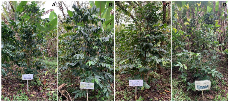
Conclusion. Under the conditions prevailing at the experimental site of the Colegio de Postgraduados Campus Córdoba, 11 C. arabica materials showed a favorable response to the severity of Hemileia vastatrix (H1 Centroamericano, Parainema, Catigua MG2, Col 1, EC 15, IPR 103, Kartila 1, Lempira, Paraíso, S795, and the Arabusta SLN 5B). This contributes to the options available to coffee growers in terms of production and plant health management against coffee leaf rust.
Share this link via
Or copy link
byÁngela Paulina Arce Leal, Edgar Antonio Rodríguez Negrete, Manuel Arturo Méndez De León, Enrique Alejandro Guevara Rivera, Norma Elena Leyva López, Jesús Méndez Lozano*
Received: 14/April/2026 – Published: 05/August/2026
DOI: https://doi.org/10.18781/R.MEX.FIT.2604-3
Abstract Background/Objective. Cucurbits are an economic pillar of global and national agriculture. However, their productivity is threatened by the increasing incidence of viral plant diseases. Consequently, coinfections with members of the Begomovirus and Crinivirus genera have been associated with exacerbated symptoms and high incidence rates in crops, leading to emerging disease patterns. The aim of this study was to identify and molecularly characterize the begomoviruses and criniviruses associated with an emerging disease in melon crops in a municipality of the Comarca Lagunera region, Mexico.
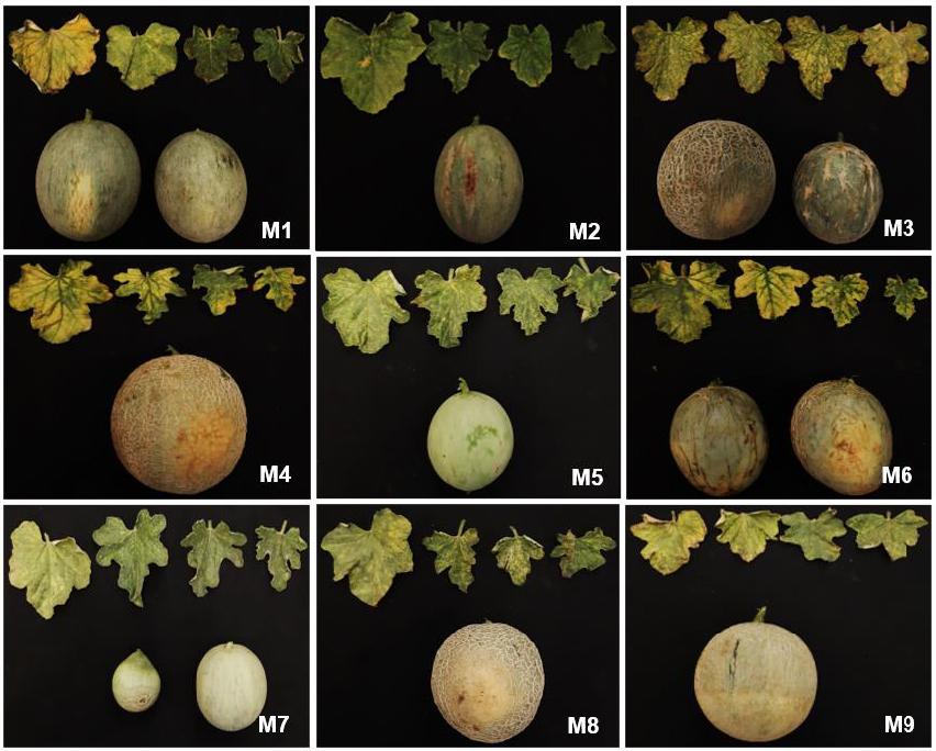
Materials and Methods. A targeted sampling of nine melon plants exhibiting symptoms such as severe interveinal chlorosis, mottling, yellow mosaic patterns, and fruit deformation was carried out. For viral detection, DNA extraction was performed using 3% CTAB and RNA extraction using the TRIzol reagent. Begomoviruses and criniviruses were detected using PCR and RT-PCR with specific primers. Complete begomovirus genomes were enriched by rolling circle amplification (RCA). Amplicons derived from Open Reading Frame (ORF) of the capsid protein (CP) of criniviruses were generated using RT-PCR. Subsequently, the genomes and CP amplicons were cloned and sequenced for phylogenetic and evolutionary analysis.
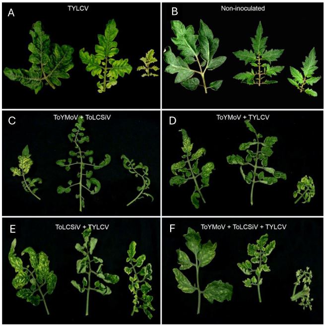
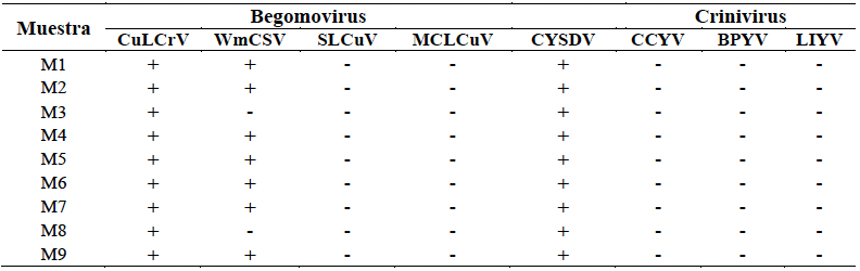
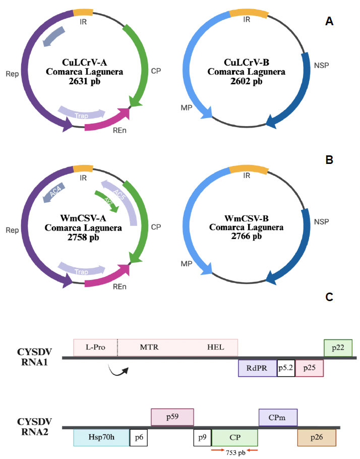
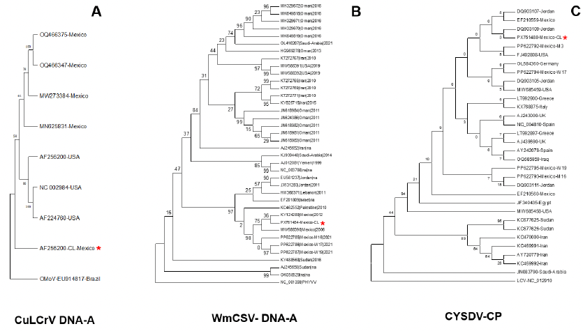
Results. Molecular identification revealed the prevalence of mixed infections in seven of nine samples, comprised of two begomovirus species, cucurbit leaf crumple virus (CuLCrV, Begomovirus cucurbitae) and watermelon chlorotic stunt virus (WmCSV, Begomovirus citrulli), and the crinivirus cucurbit yellow stunting disorder virus (CYSDV, Crinivirus cucurbitae). Genomic analyses confirmed that CuLCrV and WmCSV possess genomic structure typical of begomoviruses. Significant divergence was observed at specific loci (CuLCrV C4 gene, WmCSV NSP gene, and DNA-B intergenic regions of both species), suggesting their potential involvement in evolutionary adaptation processes. Phylogenetically, although the CuLCrV isolate showed close relationship to isolates from Arizona and Sonora, it was placed in a clade distinct from those previously reported in the region, indicating an independent evolutionary course. Furthermore, phylogenetic analysis based on the nucleotide sequence of the CYSDV capsid protein (CP) gene revealed a close relationship with isolates previously reported in Jordan and Mexico.
Conclusions. This study represents the first report in Mexico of a disease associated with WmCSV, CuLCrV, and CYSDV coinfection in cucurbits. The findings amphasize the complexity of emerging diseases and highlight the importance of strengthening molecular epidemiological surveillance to mitigate the impact of these mixed infections on Mexican agriculture.
Share this link via
Or copy link
byJatsiris A Meza Silva, Jeiry Toribio Jiménez, Renato León Rodríguez, Nelda Xanath Martínez Galero, Erubiel Toledo Hernández, Roxana Reyes Ríos, Alberto Patricio Hernández*
Received: 15/May/2026 – Published: 29/July/2026
DOI: https://doi.org/10.18781/R.MEX.FIT.2605-2
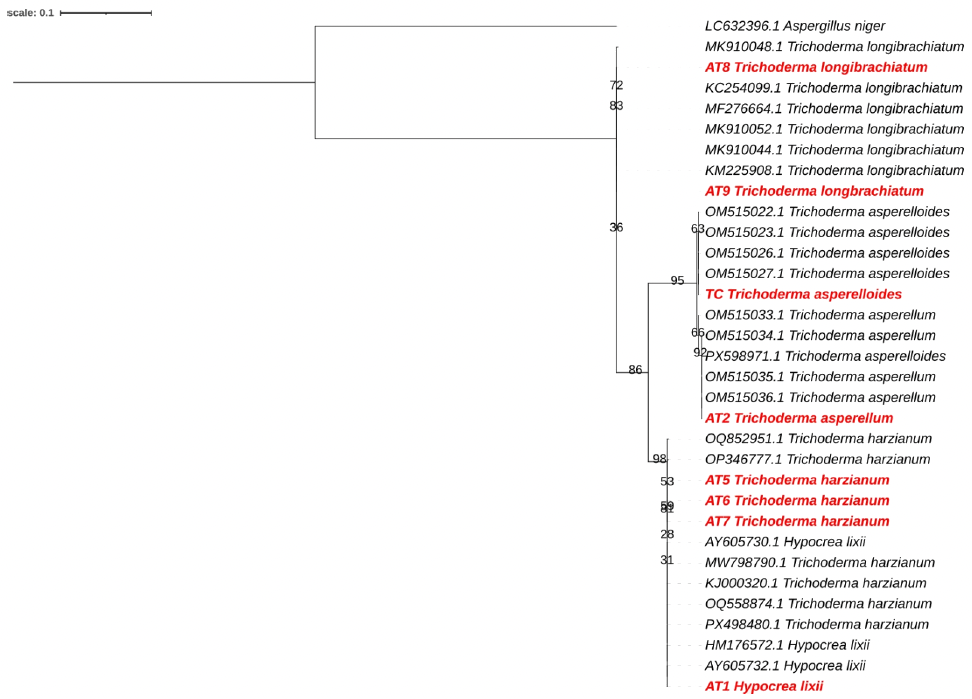
Abstract Background/Objective. The genus Trichoderma comprises fungi widely recognized for their capacity as biological control agents and plant growth promoters; however, information on their molecular diversity in agricultural systems in Guerrero, Mexico, is limited. The objective of this study was to isolate and molecularly identify native Trichoderma species present in different sites within the municipality of Chilpancingo de los Bravo, Guerrero, and to analyze their distribution in relation to management conditions.
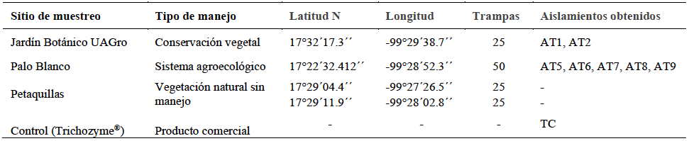
Experimental development. During August and September 2025, 125 traps with sterile rice were installed at four sampling points: Palo Blanco (Agroecological system), the Botanical Garden of the Universidad Autónoma de Guerrero (Plant conservation), and two areas of Petaquillas (Unmanaged natural vegetation). Isolates were purified by monosporic culture, morphologically characterized, and identified by ITS region sequencing and maximum likelihood phylogenetic analysis with 1,000 bootstrap replicates.
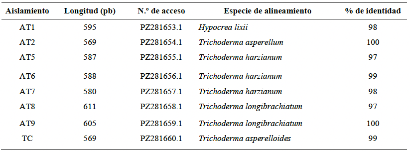
Results. Seven Trichoderma isolates were obtained, recovered only from the Palo Blanco agroecological plot and the Botanical Garden of the Universidad Autónoma de Guerrero. Phylogenetic analysis identified T. harzianum/Hypocrea lixii, T. asperellum, and T. longibrachiatum. Representatives of T. harzianum/H. lixii and T. longibrachiatum were identified at Palo Blanco, while T. harzianum/H. lixii and T. asperellum were identified at the Botanical Garden. The T. harzianum/H. lixii complex was the predominant taxon, representing 57.1% of the isolates obtained.
Conclusion. Three Trichoderma taxa were identified, with the T. harzianum/Hypocrea lixii complex predominating (57.1%), present in Palo Blanco and the Botanical Garden. The results expand our knowledge of its diversity and distribution in Guerrero, although its biotechnological potential should be confirmed through functional evaluations.
Share this link via
Or copy link
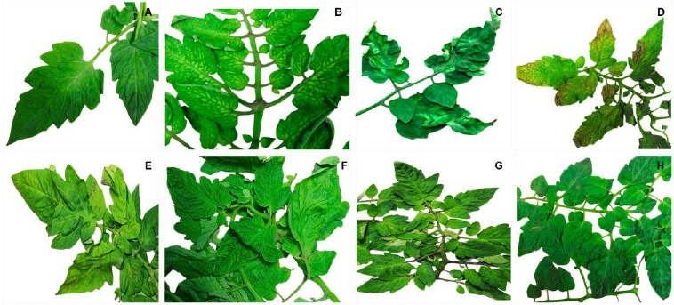
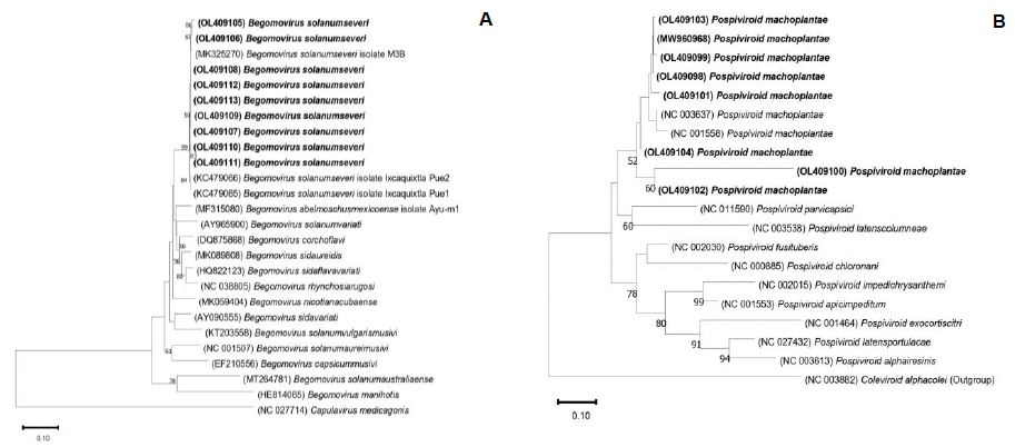
Natural co-infection of Pospiviroid machoplantae and Begomovirus solanumseveri in tomato
byMaría del Carmen Zuñiga Romano, Daniel Leobardo Ochoa Martínez*, Reyna Isabel Rojas Martínez, Sergio Aranda Ocampo, Candelario Ortega Acosta, Erika Janet Zamora Macorra
Received: 08/March/2026 – Published: 27/July/2026
DOI: https://doi.org/10.18781/R.MEX.FIT.2603-1
Abstract Background/Objective. High whitefly populations and incidence of up to 90% of tomato plants showing mosaic, intervenial chlorosis, deformation, rugosity and leaf necrosis, associated with virus and viroids, were observed at a high-tech greenhouse in Atlacomulco, State of Mexico. To identify the viruses or viroids associated with these symptoms, the withefly species, as well as to determine if the insects were carrying any of these pathogens, monthly sampling was conducted over a two-year period, collecting plant leaf tissue and adult whiteflies.
Materials y Methods. A total of 347 plants (211 symptomatic and 136 asymptomatic) were analyzed using immunostrips for impatiens necrotic spot virus (Orthotospovirus impatiensnecromaculae), tomato spotted wilt virus (O. tomatomaculae), tobacco mosaic virus (Tobamovirus tabaci), tomato mosaic virus (T. tomatotessellati), pepino mosaic virus (Potexvirus pepini) and cucumber mosaic virus (Cucumovirus CMV), as well as by RT-PCR and PCR using universal primers for viroids and viruses of the genera Pospiviroid and Begomovirus, respectively. DNA was extracted from the insects and analyzed by PCR with specific primers that amplify a fragment of the mitocondrial gene (mtCOI) to identify the whitefly species, as well as universal primers for begomoviruses to determine if the insects were carriers of these viruses.
Results. The results obtained using immunostrips were negative. The fragments amplified by RT-PCR and PCR were sequenced and corresponded to tomato planta macho viroid (Pospiviroid machoplantae) and tomato severe leaf curl virus (Begomovirus solanumseveri). Of the plants analyzed, 49.56% tested positive for the pospiviroid, 23.9% for the begomovirus, and 8.93% for both. Infection caused solely by the viroid was the only one that resulted in an asymptomatic condition. Bemisia tabaci was identified as the whitefly species and was determined to be a carrier of B. solanumseveri.
Conclusions. Symptoms of mosaic, intervenial chlorosis, leaf deformation and necrosis, blistering, and rugosity observed in tomato plants were associated with individual infection by tomato planta macho viroid (Pospiviroid machoplantae), and tomato severe leaf curl virus (Begomovirus solanumseveri), as well as co-infection by both. This is the first report of natural co-infection of tomato planta macho viroid and tomato leaf curl virus. The presence of asymptomatic plants is a key factor to consider when carrying out cultural practices to minimize mechanical transmission of the viroid.
Share this link via
Or copy link
Detection of Closterovirus tristezae in Persian lime using machine learning and drones
byMaribel Reyes García, Remigio Anastacio Guzmán Plazola*, Reyna Isabel Rojas Martínez, Victoria Ayala Escobar, Héctor Flores Magdaleno
Received: 03/October/2025 – Published: 17/July/2026
DOI: https://doi.org/10.18781/R.MEX.FIT.2510-2
Abstract Background/Objective. Persian lime (Citrus latifolia) production in Mexico is threatened by various diseases. Among the most significant is the citrus tristeza virus (Closterovirus tristezae, CTV), which can infect plants asymptomatically. This research aimed to evaluate the use of drone-acquired multispectral imagery and machine learning algorithms to identify diseased plants as a tool for early, non-destructive detection.
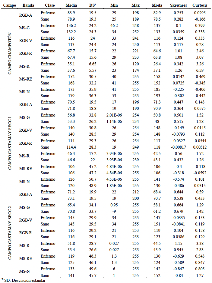
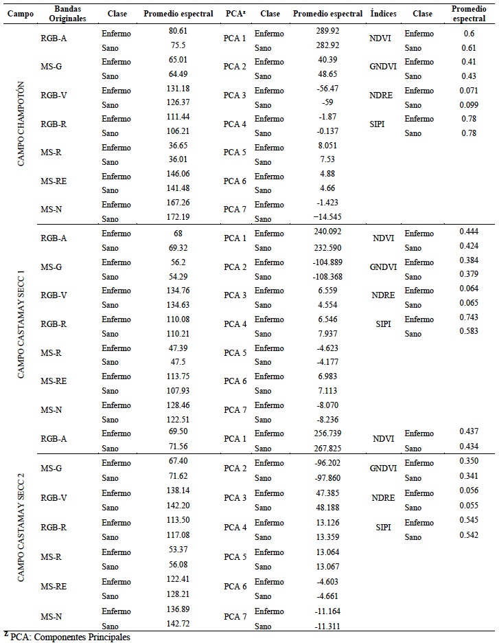
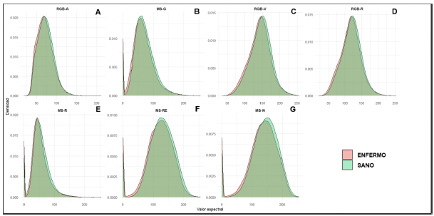
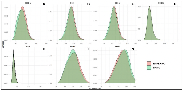
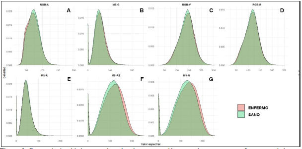
Materials and Methods. The study was conducted in three agricultural fields in Campeche, México: Champotón and Castamay (sections 1 and 2). Multispectral images (green, red, red-edge, and near-infrared) were acquired, and spectral signatures, vegetation indices (NDVI, GNDVI, NDRE, SIPI), and principal components (PCA) were extracted. Supervised classifiers—Random Forest (RF), Maximum Likelihood (MAXLIKE), Support Vector Machine (SVM), and Multilayer Perceptron (MLP)—were applied using hard and soft classification schemes.
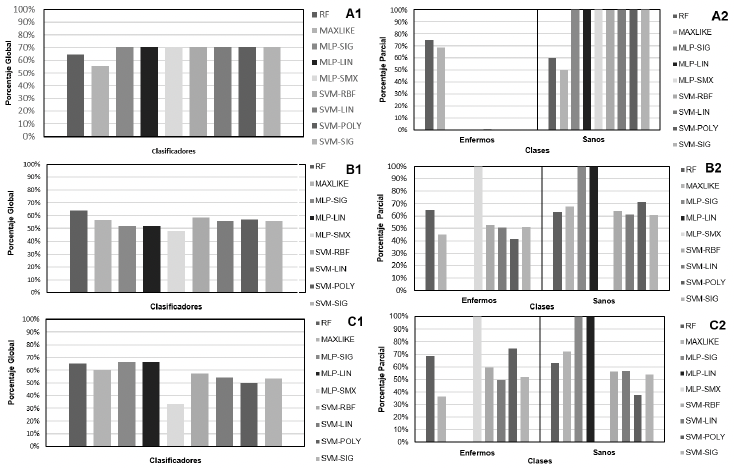
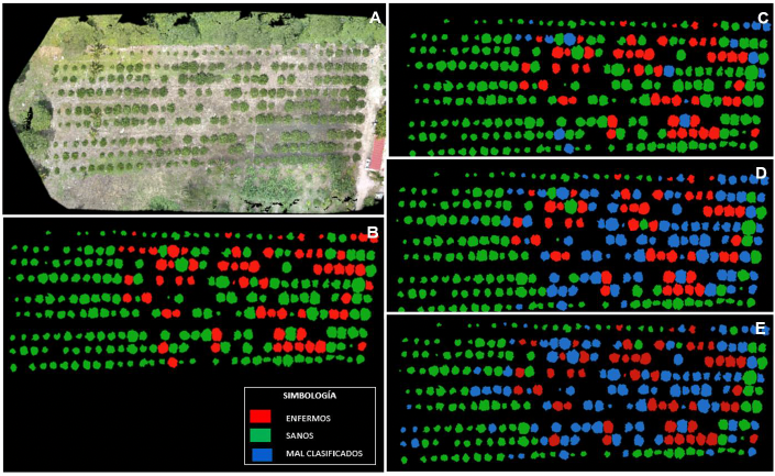
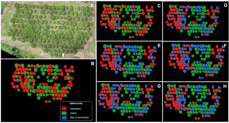
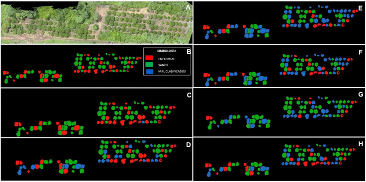
Results. RF performed the best. In hard classification, it achieved overall accuracies of 64% in Champotón and Castamay Section 1, and 65% in Section 2, with diseased plant detection rates between 65 and 75%. It yielded better results in soft classification. In Champotón, it achieved 94% overall accuracy and detected 79% of diseased plants. In Castamay Section 1, the figures were 91 and 92%, respectively; and in Section 2, it achieved 99% overall accuracy and 100% disease detection.
Conclusion. Random Forest demonstrated the most robust and stable performance across the various scenarios and fields analyzed—in both hard classification (65%) and soft classification (94%)—confirming its ability to handle non-linear relationships and spectral variability in real agricultural environments. The use of multispectral imagery combined with machine learning algorithms—specifically Random Forest (RF) for soft classification—represents an effective tool for early diagnosis of CTV in Persian lime.
Share this link via
Or copy link
byJesús Eduardo Ramírez Méndez, Meralida Bartolón Pérez, Francisco Daniel Hernández Castillo, Yisa María Ochoa Fuentes, Laura Castro Rosalez, Marco Antonio Tucuch Pérez*
Received: 19/January/2026 – Published: 13/July/2026
DOI: https://doi.org/10.18781/R.MEX.FIT.2601-3
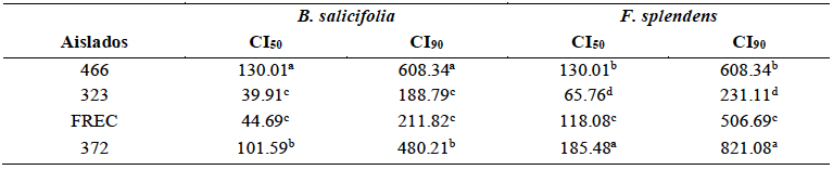
Abstract Background/Objective. Neopestalotiopsis rosae is the causal agent of crown and root rot in strawberry, resulting in significant yield losses. The aim of this study was to evaluate the sensitivity of four N. rosae isolates (466, 323, FREC, and 372) to plant extracts (PEs) obtained from Baccharis salicifolia and Fouquieria splendens, and to determine their in vitro antifungal activity.
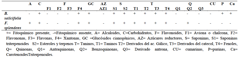
Experimental development. A qualitative phytochemical screening was performed to identify the major bioactive compounds present in the PEs. Antifungal activity was evaluated using in vitro microdilution assays at concentrations ranging from 3.9 to 2 000 mg L⁻¹ (10 concentrations). The 50 and 90% inhibitory concentrations (IC₅₀ and IC₉₀) were estimated, and the data were analyzed using analysis of variance (ANOVA) followed by Tukey's multiple comparison test (p ≤ 0.05). The effects of the PEs on fungal cellular structures were also assessed.
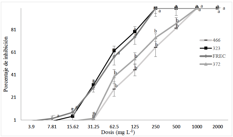
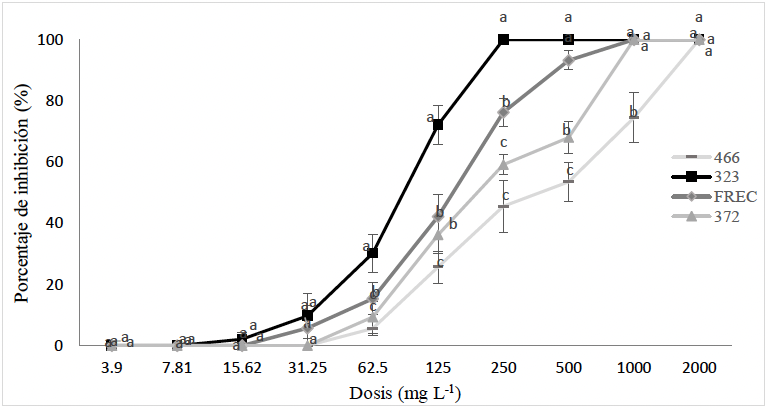
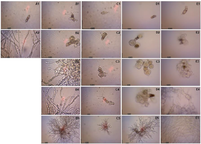
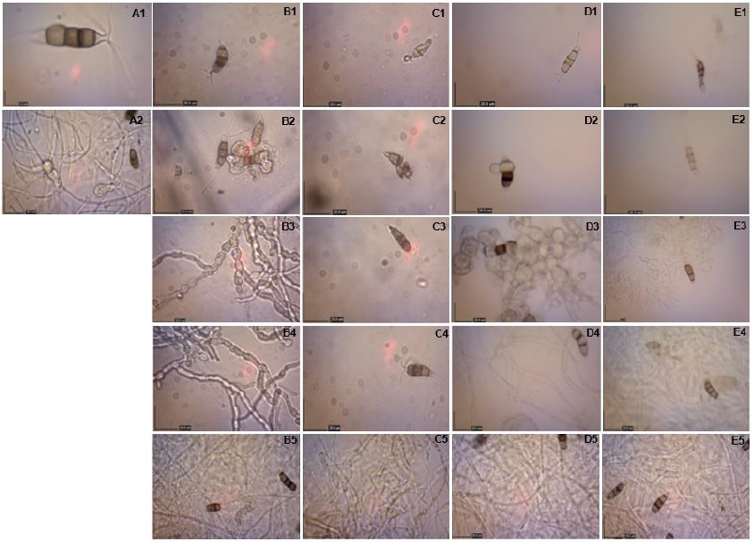
Results. Phytochemical screening revealed the presence of phenolic compounds, saponins, and tannins in both plant extracts. Among the N. rosae isolates, isolates 323 and FREC were the most sensitive, exhibiting complete inhibition with both PEs at 250 mg L⁻¹. In contrast, isolates 372 and 466 required higher concentrations to achieve complete inhibition. B. salicifolia was more effective against isolates 466 and 372, achieving complete inhibition at 1 000 mg L⁻¹, whereas F. splendens required 2 000 mg L⁻¹ to completely inhibit isolate 466. Structural analyses revealed that isolate 323 was most susceptible to extract-induced damage at 250 mg L⁻¹ and above, as evidenced by inhibition of conidial germination and severe morphological alterations, including cellular collapse and dehydration. Isolates FREC and 372 exhibited mycelial thickening at 500 mg L⁻¹, whereas isolate 466 was the least sensitive, showing no evident structural damage even at this concentration.
Conclusions. Plant extracts from B. salicifolia and F. splendens contain bioactive phytochemicals with antifungal activity against N. rosae. Isolates 323 and FREC were the most sensitive, exhibiting 100% inhibition and pronounced structural damage at 250 mg L⁻¹, highlighting the potential of these extracts as biorational alternatives for the management of N. rosae. Nevertheless, further greenhouse and field studies are required to validate their efficacy under commercial production conditions.
Share this link via
Or copy link
bySantiago Vergara Pineda, Paola S Puga Guzmán, Daniel Mendoza Jiménez, José Antonio Cervantes Chávez*
Received: 09/March/2026 – Published: 22/June/2026
DOI: https://doi.org/10.18781/R.MEX.FIT.2603-2
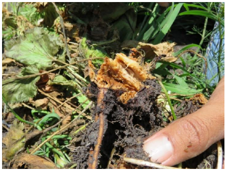
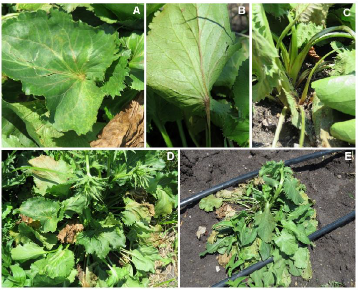
Abstract Background/Objective. Plant species of the genus Eryngium are native to Mexico; their cultivation as ornamental cut flowers is a recent development, so the pathogens that affect them are largely unknown. In this context, a grower set out to innovate by using this plant as a cut flower but encountered a rot disease that decimated his field; therefore, the objective of this study was to isolate, identify, and characterize the causal agent of soft rot in the stems of Eryngium spp.
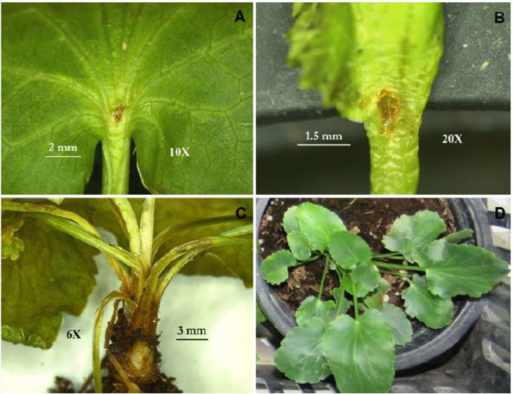
Experimental development. Plants exhibiting rot symptoms were collected from a cultivated plot in the municipality of Ixtapan de la Sal, State of Mexico. From ten symptomatic plants kept in a humid chamber, a bacterium was isolated and identified using biochemical analysis. For molecular identification, DNA was extracted, and the 16S ribosomal gene was amplified with primers 27F and 1492R. PCR products were sent to the Advanced Genomics Laboratory at LANGEBIO, CINVESTAV Irapuato, Guanajuato. Koch’s postulates were also performed on twenty asymptomatic Eryngium spp. seedlings, which were inoculated by introducing a bacterial mass at the leaf base, the distal petiole, and the basal vein region.
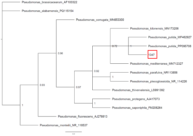
Results. Based on biochemical analysis, the bacterium was identified as belonging to the genus Pseudomonas; molecular analysis revealed that the bacterium was Pseudomonas putida. According to Koch’s postulates, 100% of the symptoms associated with soft rot were observed.
Conclusion. Pseudomonas putida was confirmed as the causal agent of soft rot in Eryngium spp. Its pathogenicity was confirmed in Eryngium spp. seedlings according to Koch's postulates. This is the first report of P. putida causing this disease in these plants.
Share this link via
Or copy link