

Share this link via
Or copy link

Article filters
Search Papers:
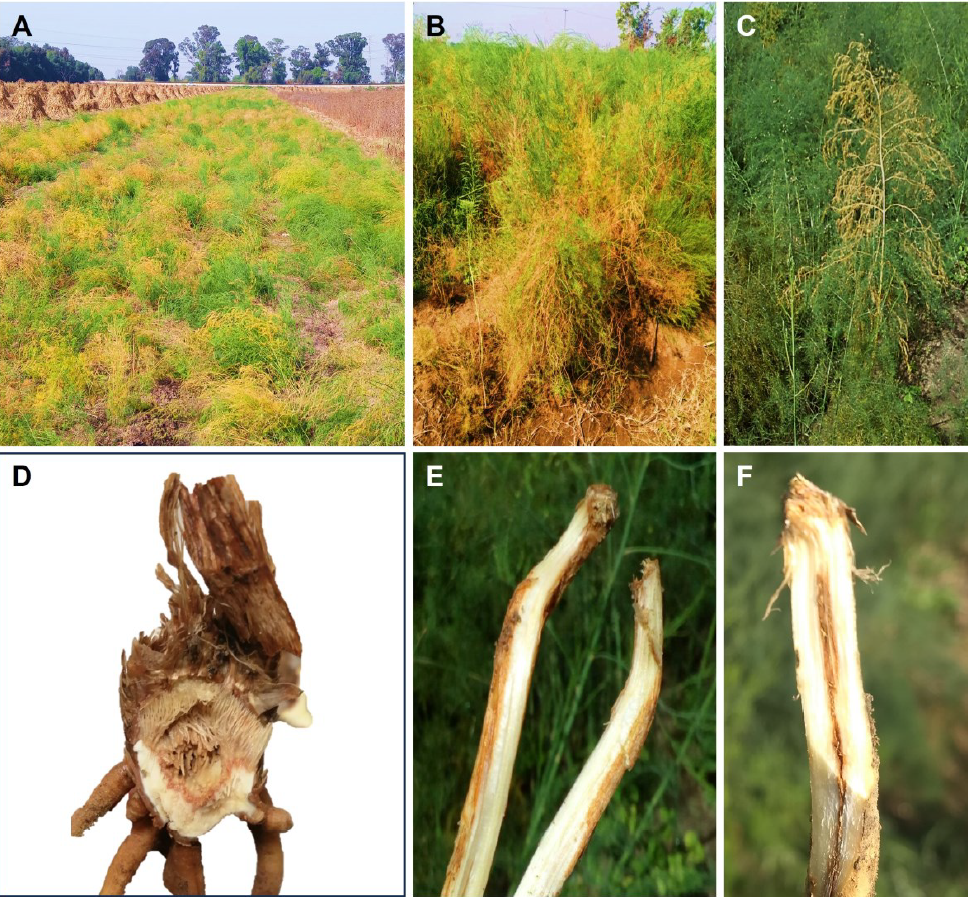
Etiology of rhizome rot of asparagus (Asparagus officinalis) in Atenco, Mexico State
byJuan Agustin Gonzalez Cruces*, José Sergio Sandoval Islas, Cristian Nava Díaz, Maricarmen Sandoval Sánchez
Received: 30/November/2023 – Published: 16/November/2023
DOI: https://doi.org/10.18781/R.MEX.FIT.2211-1
Abstract Background/Objective. The objective of this research was to identify the causal agent of asparagus rhizome rot, as well as evaluate different inoculation methods and the severity of the isolates.
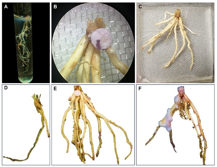
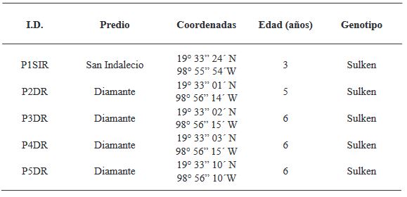
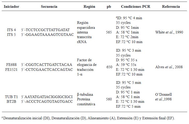
Materials and Methods. Sampling was carried out in five producing plots Atenco, Edo. from Mexico. Five isolates of Fusarium spp. were selected. (one per plot) to perform pathogenicity tests. Three isolates were selected for their colonization characteristics for severity tests with different inoculation methods: Immersion for 12 h, immersion for 30 min and inoculation by contact with absorbent paper soaked in 1 mL of inoculum. Concentrations of 1x10⁶ conidia mL-1 were used. 10 rhizomes were used per treatment and 10 rhizomes without inoculation. To determine the severity, photographs (in GIMP®) of the rhizome were analyzed seven days after inoculation. The isolates were molecularly identified with ITS4/ ITS5, EF688/EF1521 and TUBT1/BT2B.
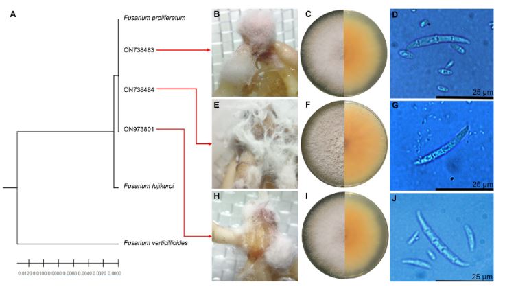
Results. Fusarium prolifetatum was morphologically and molecularly identified in the three isolates. The P3DR isolate was the most severe (14.6%), followed by P5DR (13.9%) and P1SIR (11.6%).
Conclusion. The most effective inoculation method was immersion for 30 min. They were registered in the NCBI Gene Bank with accessions ON738484 (P3DR), ON973801 (P5DR) and ON738483 (P1SIR). This is the first report of F. prolifetatum in the Edo. from Mexico.
Share this link via
Or copy link

Diagrammatic scale to evaluate the severity of gray mold (Botrytis cinerea) in pomegranate
byAlberto Patricio Hernández, Yuridia Mercado Flores*, Alejandro Téllez Jurado, María del Rocío Ramírez Vargas, Andrés Quezada Salinas
Received: 30/October/2022 – Published: 15/June/2023
DOI: https://doi.org/10.18781/R.MEX.FIT.2302-9
Abstract The aim of this study was to design and validate a diagrammatic scale to estimate the severity of gray mold induced by Botrytis cinerea in pomegranate cultivation. A total of 120 healthy and diseased fruits with varying degrees of affliction were collected from orchards with active production located in the municipalities of Chilcuahutla and Taxquillo in the state of Hidalgo, Mexico (20° 18’ 11’’ N, 99° 14’ 23’’ W, 20° 32’ 01’’ N, 99° 20’ 03’’ W, respectively). From these, 60 were selected to determine the severity percentage, according to a 6-class scale (Class 0 = 0%, Class 1 = >0% - 5% - 10%, Class 2 = >10% - 25% - 50%, Class 3 = >50% - 75% - 85%, Class 4 = >85% - 90% - 95%, and Class 5 = >95% - 100%), using the 2LOG software. With the obtained data, representative images were selected to build the diagrammatic scale using Adobe Photoshop. The accuracy (r2), precision (β0), and reproducibility (β1) were verified by simple linear regression applied to the data obtained by 12 evaluators with and without experience in the observation of plant diseases. As a result, values of r2 of 0.42 and 0.85 were obtained, without and with the use of the scale, respectively, which confirmed that this tool is suitable to evaluate the severity of the disease accurately and reproducibly.
Share this link via
Or copy link
There are no images, at the moment, loaded for this article.
In vitro antagonism of Trichoderma against Rhizoctonia solani
byJesús Orlando Pérez González, Sergio Gavino Ramírez Rojas, Ramiro Rocha Rodríguez, Katya Ornelas Ocampo, Jorge Miguel Vázquez Alvarado, Filogonio Jesús Hernández Guzmán, Mariel Garduño Audelo*
Received: 30/October/2022 – Published: 15/June/2023
DOI: https://doi.org/10.18781/R.MEX.FIT.2304-2
Abstract Trichoderma spp., is a highly efficient antagonist of root pathogens, such as Rhizoctonia solani, which causes loss in many crops. The aim of this research was to evaluate in vitro the antagonistic capacity of T. viride, T. koningii, T. harzianum and Trichoderma spp. isolates against R. solani from a potato crop. In confrontation tests, all Trichoderma isolates were classified as antagonists class 2 according to Bell scale, where T. harzianum and T. koningii showed more than 60% inhibition of the radial growth of R. solani at 120 h. In the interaction between T. harzianum and Trichoderma spp. with R. solani, as mycoparasitism strategy, vacuolization, lysis, coiling, and penetration were demonstrated, the last two were present in all Trichoderma isolates evaluated.
Share this link via
Or copy link
There are no images, at the moment, loaded for this article.
byOsvaldo Jhosimar Couoh Dzul, Karla Gisel Carreón Anguiano, Blondy Canto Canché*
Received: 30/October/2022 – Published: 15/June/2023
DOI: https://doi.org/10.18781/R.MEX.FIT.2210-5
Abstract The control of phytopathogens is key for food security. In the last decade, the use of interference RNA (iRNA) has been proposed as a technological tool for controlling diseases and pests in agriculture. Although different approaches have been described, such as the use of “Host-Induce Gene Silencing” (HIGS) and “Virus-Induce Gene Silencing” (VIGS), more recently a non-transgenic and environmentally friendly approach has emerged, called “Spray -Induce Gene Silencing” (SIGS), which uses double-stranded “naked” RNA (dsRNA). This review discusses recent reports on the use of dsRNA, especially SIGS, to control phytopathogenic fungi; emphasizing factors such as efficacy, safety in terms of human health and its stability in the environment. It also focuses on important phytosanitary problems in Mexico and Latin America that can be addressed with SIGS. This review concludes that SIGS technology has real potential to be used to control phytopathogenic fungi on plants in the field and on postharvest fruits. At the end, the critical tasks and the lines of research that must be carried out to promote the SIGS to make it a reality are considered.
Share this link via
Or copy link
There are no images, at the moment, loaded for this article.
byEdna Esquivel Miguel, José Luciano Morales García*, Martha Elena Pedraza Santos, Ana Tztzqui Chávez Bárcenas, Soledad García Morales, Samuel Pineda Guillermo
Received: 01/May/2023 – Published: 24/August/2023
DOI: https://doi.org/10.18781/R.MEX.FIT.2302-3
Abstract Elsinoe perseae (= Sphaceloma perseae) is the causal agent of the Mancha purpura or scab in avocado (Persea americana). In this study, the pathogenicity and virulence of E. perseae isolates from different agroecologicalproducing areas of Michoacán, Mexico will be reduced. For this, nursery plants with avocado fruits of Flor de María and Méndez varieties were used. On the other hand, the in vitro sensitivity of chemical fungicides (Azoxystrobin, thiabendazole, Pyraclostrobin, Cyprodinil + Fludioxonil and Azoxystrobin + Propiconazole) and authorized for use in orchards with organic management (copper sulfate, copper gluconate, copper oxychloride and the plant extract Larrea tridentata). The observed symptoms of Mancha purpura in the inoculated fruits were corroborated with those described for E. perseae in avocado. Inoculated fruits Flor de María variety shows the highest susceptibility to the pathogen. The isolates of E. perseae presented different degrees of virulence. The isolates showed different in vitro sensitivity values to the fungicides evaluated in the experiment. The pathogen showed the most sensitivity in vitro to chemical fungicides: thiabendazole and Azoxystrobin + Propiconazole (100% inhibition), and to those authorized in orchards with organic management: L. tridentata and copper oxychloride (on average 58% inhibition).
Share this link via
Or copy link
There are no images, at the moment, loaded for this article.
Effect of adjuvants, fungicides and insecticides on the growth of Trichoderma koningiopsis Th003
byYimmy Alexander Zapata Narváez*, Blanca Lucia Botina Azain
Received: 01/May/2023 – Published: 24/August/2023
DOI: https://doi.org/10.18781/R.MEX.FIT.2305-1
Abstract The effect of 44 agrochemicals (fungicides, insecticides and adjuvants) on the mycelial growth and germination conidia of Trichoderma koningiopsis Th003 was determined by seeding of 5 mm discs of fungal mycelium in Sabouraud agar supplemented with each agrochemical and seeding of conidia suspended in solutions of agrochemicals in water agar. For the adjuvants, their effect on the phyllospheric establishment of T. koningiopsis Th003 was determined by following their population in cape gooseberry leaflets inoculated with the fungus suspended in them. Eight fungicides did not inhibit the fungus mycelial growth or the conidia germination. Fenhexamid - Tebuconazole, Flutriafol and Kasugamicina inhibited it between 34 and 48% without affecting germination, Thiram - Pyrimethanil, Prochloraz, Tiabendazol, Spiroxamina and Triadimenol - Tebuconazole inhibited the growth and Thiram - Pyrimethanil and Dodine did not allow conidia germination. Insecticides and adjuvants presented an inhibition of up to 70% but did not affect the conidia germination. No negative effects of the adjuvants on the phyllosphere establishment of T. koningiopsis Th003 were observed, recovering from the treatments approximately 1x10³ CFU g-1.
Share this link via
Or copy link
There are no images, at the moment, loaded for this article.
byNidia Esther Hau Yama, Marianne Soledad Luna Rivero, Blondy Beatriz Canto Canché, Yereni Minero García, Cecilia Hernández Zepeda, Diana Sánchez Rangel, Oscar Alberto Moreno Valenzuela*
Received: 10/February/2022 – Published: 31/July/2023
DOI: https://doi.org/10.18781/R.MEX.FIT.2305-4
Abstract In the literature, RNA-based interference methods have been proposed for the control of pathogenic fungi in plants, such as virus-mediated silencing (VIGS). The objective of this work was to evaluate the functionality and efficiency of the VIGS EuMV-YP∆CHS4 vector, which contains a fragment of the Fusarium sp. chitin synthase 4 (CHS4) gene sequence, and was constructed from the EuMV-YP vector:∆AV1. The VIGS vector was evaluated under two conditions in Nicotiana benthamiana plants previously inoculated with Fusarium sp. and in plants with inoculation after the inoculation of the VIGS vector. The results showed that in both treatments with the EuMV-YP∆CHS4, a delay in leaf fall during the first days of infection and a decrease in the stem damage of almost one centimeter, compared to the control infected with the fungus. In addition, a reduction of up to 30% in the relative expression of the CHS4 gene was observed. These results suggest that the application of VIGS vectors may be efficient as a strategy to protect plants against Fusarium sp.
Share this link via
Or copy link
There are no images, at the moment, loaded for this article.
byLaura Belén Tapia de la Barrera, Manuel Alonzo Báez Sañudo, Raymundo Saúl García Estrada, Juan Manuel Tovar Pedraza, José Armando Carrillo Fasio
Received: 10/February/2022 – Published: 31/July/2023
DOI: https://doi.org/10.18781/R.MEX.FIT.2210-2
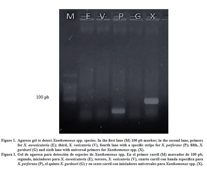
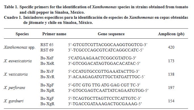
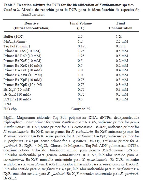
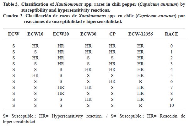
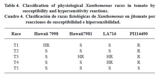
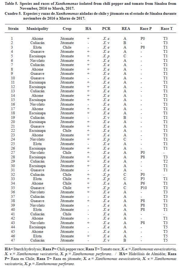
Abstract Bacterial spot of tomato and chili pepper, caused by four species of Xanthomonas and various races, is one of the diseases with the greatest impact on horticulture worldwide. The aim of this study was to identify the species and physiological races of Xanthomonas present in tomato (Solanum lycopersicum) and chili pepper (Capsicum annuum) crops in Sinaloa, Mexico. For this purpose, samples with typical symptoms of bacterial spot were collected in commercial fields of the different municipalities in the state of Sinaloa. Ninety-three bacteria were isolated on semi-selective medium as nutrient agar and yeast extract-dextrose. A total of 47 bacteria were identified as Xanthomonas by a combination of morphological, pathogenic, biochemical, physiological, and molecular tests. In addition, to characterize the morphological race of each strain, differential tomato (four) and chili pepper (six) lines were used. Molecular diagnostic using specific primers indicated that 83% corresponded to X. euvesicatoria, 10.6% to X. perforans, and 6.4% to X. vesicatoria. At the physiological race level, races T1, T2, T3, and T5 were detected in tomato; while the Xanthomonas races detected in chili pepper were P0, P3, P6, P8 and P10. This information updates previous data on the distribution of races of Xanthomonas that infect chili peppers in Sinaloa, since races P6 and P10 are reported for the first time.
Share this link via
Or copy link
byYimmy Alexander Zapata Narváez*, Andrés Díaz Garcia, Camilo Rubén Beltrán Acosta
Received: 10/February/2022 – Published: 31/July/2023
DOI: https://doi.org/10.18781/R.MEX.FIT.2302-5
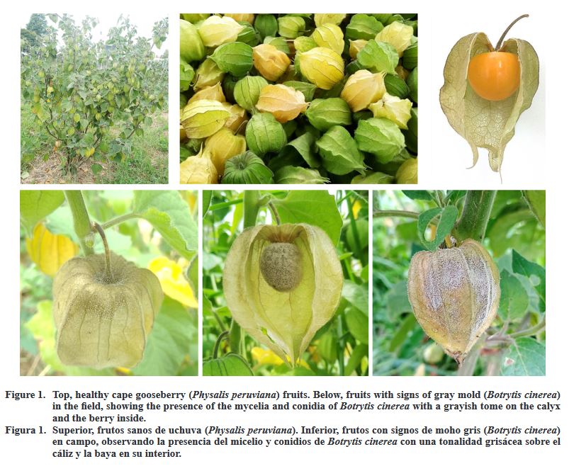
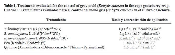
Abstract The effect of the field applications of three bioproducts (based on Trichoderma koningiopsis, Rhodotorula mucilaginosa, and Bacillus amyloliquefaciens), the alternation of the biostimulant Kendal® and the Swinglea glutinosa extract and the rotations of two fungicides (based on Azoxystrobin-Difenoconazole and Thiram- Pyrimethanol), on the incidence of gray mold in cape gooseberry postharvest was evaluated. For this purpose, the fruit was harvested weekly in the field, arranged in wet chambers with fruits with and without calyx, and incubated for seven days at 20 °C in laboratory conditions to promote the development of B. cinerea and determine the efficacy in its control. In addition, the populations of the microbial antagonists were monitored between applications by collecting the leaflets and washing them in 0.1% Tween 80 and sowing aliquots in specific culture media. In fruits with calyx, the lowest incidence of the gray mold, with averages of 48 and 51%, occurred with the applications of the bioproducts based on T. koningiopsis and R. mucilaginosa, respectively. In contrast, the incidence did not exceed 1.4% in fruits without calyx in all treatments. Furthermore, the population of microbial antagonists in the phyllosphere remained constant between applications, with counts of 1x10³ CFU g-1 for T. koningiopsis and 1x10⁵ CFU g-1 for R. mucilaginosa and B. amyloliquefaciens.
Share this link via
Or copy link
byWendy Abril Flores Pérez, Nemesio Villa Ruano, Efraín Rubio Rosas*
Received: 31/January/2022 – Published: 23/August/2023
DOI: https://doi.org/10.18781/R.MEX.FIT.2303-4
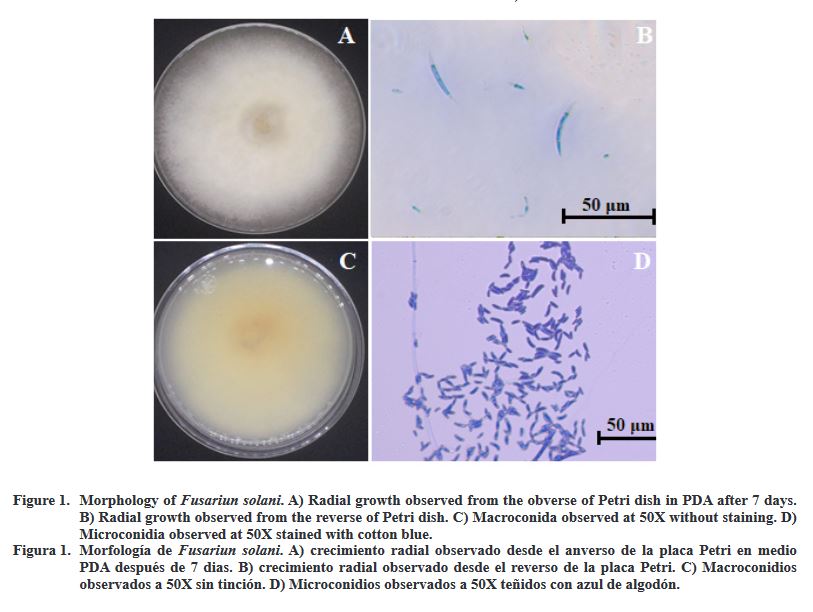
Abstract Little is known on the use of biodegradable films for the control of diseases in crops of agricultural importance. Consequently, the objective of this work was to determine the efficacy of chitosan hybrid films impregnated with thyme essential oil on Hass avocado fruits previously infected with Fusarium solani isolated from the northeastern highlands of Puebla, Mexico. The native strain of F. solani was morphologically and molecularly identified and the fungistatic activity of three chitosan films supplemented with 0.7% (FT1), 1.0% (FT2) and 1.3% (FT3) of thyme essential oil was evaluated in situ on the growth of F. solani in avocado fruits. Texture, transmittance and opacity of the films were obtained by scanning electron microscopy and UV-Vis spectrometry, respectively. Overall, it was recorded that films with a higher concentration of thyme essential oil (1-1.3% w/v) presented lower transmittance in the UV light range and higher opacity. Avocado fruits infected with F. solani simultaneously treated with FT2 and FT3 reduced the appearance of symptoms while preserved firmness, as well as fiber, fat, reducing sugars, and protein content (p < 0.01). In the same context, these materials promoted the conservation of the content of the nutraceuticals linoleic acid, oleic acid, palmitic acid and palmitoleic acid for 21 days. These results suggest that the hybrid films generated in the present study have the ability to control fusariosis caused by this fungus, prolonging the shelf life of Hass avocado fruit.
Share this link via
Or copy link