RMF Logo

Article filters

Search Papers:

For searching before 2024 see Archive (2000-2023)

All the Articles

  • Vol. 2025, 43(4)
  • Review Article
  • Special Issue

Agroecological alternatives for disease management in Capsicum crops

byWendy Abril Coyotl Pérez, Mariana Flores Hernández, Cesar Agustín Ramírez Díaz, Nemesio Villa Ruano*

Received: 23/November/2024 – Published: 07/October/2025
DOI: https://doi.org/10.18781/R.MEX.FIT.2024-20

Abstract Justification. Capsicum spp. fruits are widely recognized and marketed worldwide due to their relevance in gastronomy and the pharmaceutical industry. The five most important species are C. annuum, C. baccatum, C. chinense, C. frutescens and C. pubescens; however, more than 43 wild species have been reported to date. Pepper plants and fruits are susceptible to diseases caused by phyopathogenic bacteria and fungi which are routinely controlled with synthetic products. These compounds generate serious side effects for both the environment and human health. Similarly, their use promotes increased microbial resistance and more severe diseases in crops. For this reason, the present review addresses the main diseases affecting pepper crops as well agroecological alternatives for controlling using antagonistic microorganisms and plant-based bioproducts to reduce the incidence and severity of diseases.

Theoretical and Experimental framework. An updated literature review was conduced regarding the diseases affecting different Capsicum species, as well as the agroecological alternatives available for their management.

Conclusions and Perspectives. Pepper production faces challenges due to diseases and the excessive use of agrochemicals which promote microbial resistance and environmental damage. Antagonistic microorganisms, plant extracts and essential oils offer a viable agroecological alternative to control phytopathogens without negative impacts. Although these strategies provide medium- and long-term benefits further research is required to control viral diseases and achieve sustainable production.

Show Figures and/or Tables
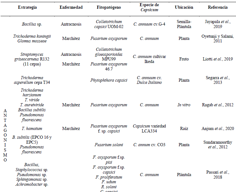
Table 1. Agroecological strategies to control major pathogens causing diseases in <em>Capsicum</em> plants.
Table 1. Agroecological strategies to control major pathogens causing diseases in Capsicum plants.
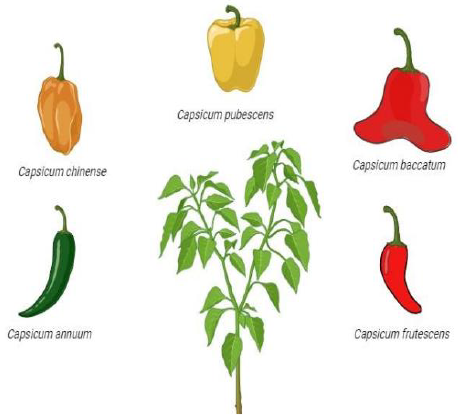
Figure 1. Fruit phenotypes of domesticated <em>Capsicum</em> species.
Figure 1. Fruit phenotypes of domesticated Capsicum species.
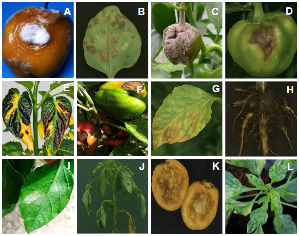
Figure 2. Symptoms of diseases caused by A) <em>Fusarium</em>, B) <em>Pythium</em>, C) <em>Botrytis</em>, D) <em>Phytophtora</em>, E) <em>Xanthomonas</em>, F) <em>Alternaria</em>, G) <em>Verticillium</em>, H) <em>Rhizoctonia</em>, I) <em>Leiveillula</em>, J) <em>Ralstonia</em>, K) <em>Erwinia</em> and L) TMV in <em>Capsicum</em> species.
Figure 2. Symptoms of diseases caused by A) Fusarium, B) Pythium, C) Botrytis, D) Phytophtora, E) Xanthomonas, F) Alternaria, G) Verticillium, H) Rhizoctonia, I) Leiveillula, J) Ralstonia, K) Erwinia and L) TMV in Capsicum species.
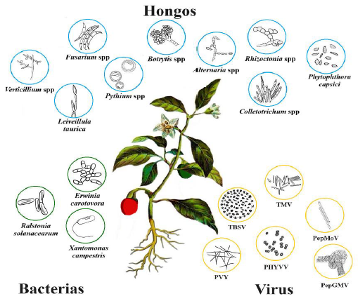
Figure 3. Main pathogens causing diseases in pepper crops.
  • Vol. 2025, 43(4)
  • Scientific Article
  • Special Issue

Effect of organic amendments on the control of Meloidogyne incognita and on the physicochemical properties of the soil

byMaría de Jesús Briseño López, Sergio Zamora Salgado, Gregorio Lucero Vega, Mirella Romero Bastidas*

Received: 30/November/2024 – Published: 03/October/2025
DOI: https://doi.org/10.18781/R.MEX.FIT.2024-29

Abstract Background/Objective. Organic amendments applied in agricultural systems commonly provide nutrients to crops. However, there are few studies on their simultaneous differential response to Meloidogyne incognita control and soil physicochemical characteristics. In this study, three organic amendments (vermicompost, cow dung, and neem green manure) were compared for root-knot nematode control and their influence on soil edaphic characteristics.

Materials and Methods. Agricultural soil, previously sterilized and mixed with the organic amendments individually, was placed in 4-L plastic pots. A treatment with a mixture of the three amendments, a synthetic fertilizer, and the water control were added and established in a randomized complete block design with 15 replicates. Each pot was inoculated with 500 juvenile M. incognita. Physicochemical parameters were analyzed 20 days later, and the nematode population and reproduction factor were determined 90 days later.

Results. The vermicompost amendment significantly influenced (P = 0.0001) the M. incognita population (≤80%) compared to the control (100%). It also significantly influenced soil variables, such as organic matter (1.11%) compared to the other treatments (0.27%).

Conclusion. The effective reduction in nematode populations was primarily recorded with vermicompost (111 nematodes/g of soil) and manure (505) amendments compared to the control (1,037). These results provide important information on the impact of the amendments on plant pathogenic nematode control and soil characteristics.

Show Figures and/or Tables
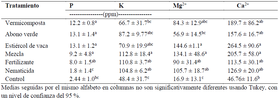
Table 1. Comparison of the total content of phosphorous and potassium, and exchangeable bases in soils treated with organic amendments.
Figure 1. Physical characterization of the soil with the different organic amendments. Error bars indicate standard deviation; different letters on the bars indicated significant differences between amendments for each parameter (Tukey test, p < 0.05).
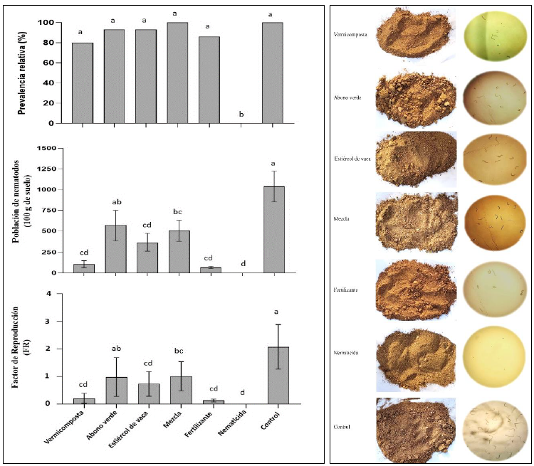
Figure 2. Relationship between the evaluated organic amendments and the population response of <em>Meloidogyne incognita</em>. Equal alphabet means indicate no significant differences among treatments according to Tukey's test, with p < 0.05.
Figure 2. Relationship between the evaluated organic amendments and the population response of Meloidogyne incognita. Equal alphabet means indicate no significant differences among treatments according to Tukey's test, with p < 0.05.
  • Vol. 2025, 43(4)
  • Phytopathological Note
  • Special Issue

Effect of biological treatments and water deficit on Macrophomina phaseolina infection in sorghum

byKarina Kuri Aguirre, Maribel Leal Castillo, Jesús Gerardo García Olivares, Sanjuana Hernández Delgado*

Received: 25/November/2024 – Published: 25/September/2025
DOI: https://doi.org/10.18781/R.MEX.FIT.2024-22

Abstract Background/Objective. Tamaulipas is the main producer of sorghum (Sorghum bicolor) in Mexico, but its unit yields are affected by biotic and abiotic factors, where Macrophomina phaseolina stands out, whose control is partially effective. In this work, the effect of biological treatments on the development of M. phaseolina in sorghum seedlings grown in a greenhouse in Reynosa, Tamaulipas, was evaluated.

Materials and Methods. Two experiments were established under a factorial arrangement in plots subdivided 2 x 2 x 5 where the large plots were the Pioneer 8282 and RB Patrón hybrids; the medium plots the levels of humidity, irrigation and water deficit; and the small plots the biological treatments: strain T808 of Trichoderma koningiopsis, strain AF36 of Aspergillus flavus; Kuali® mycotoxin chelating agent; control inoculated with M. phaseolina and absolute control.

Results. The RB-Patrón hybrid showed the greatest damage to M. phaseolina and the lowest growth (plant height: 10%) and accumulation of dry biomass (between 35-40%). Water deficit increased charcoal rot infection and reduced sorghum growth. T. koningiopsis strain T808 reduced the severity of charcoal rot (value of 4.6) while the other biological treatments exhibited severity values greater than 6.0.

Conclusion. The treatments with better sorghum growth and less damage by M. phaseolina (T808, P-8282) should be evaluated under field conditions to confirm the viability of their application in commercial sorghum production in northern Tamaulipas.

Show Figures and/or Tables
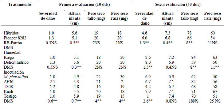
Table 1. Means comparison of the main effects for the severity of damage by <em>M. phaseolina</em>, plant height and accumulation of biomass in sorghum. First and sixth evaluations in experiment 1.
Table 1. Means comparison of the main effects for the severity of damage by M. phaseolina, plant height and accumulation of biomass in sorghum. First and sixth evaluations in experiment 1.
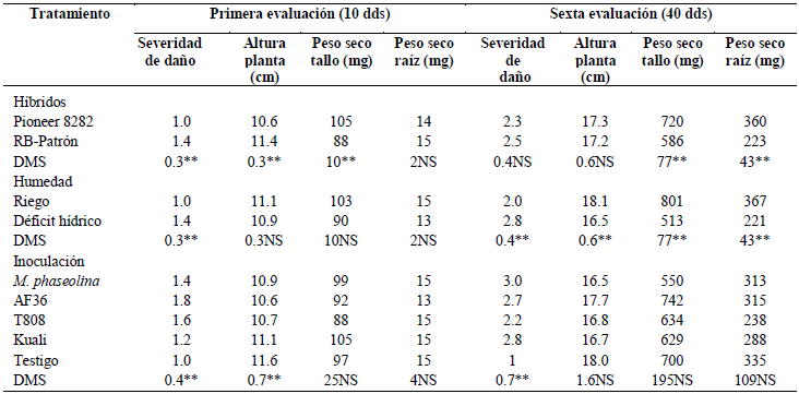
Table 2. Means comparison of the main effects for the severity of damage by <em>M. phaseolina</em>, plant height and accumulation of biomass in sorghum. First and sixth evaluations in experiment 2.
Table 2. Means comparison of the main effects for the severity of damage by M. phaseolina, plant height and accumulation of biomass in sorghum. First and sixth evaluations in experiment 2.
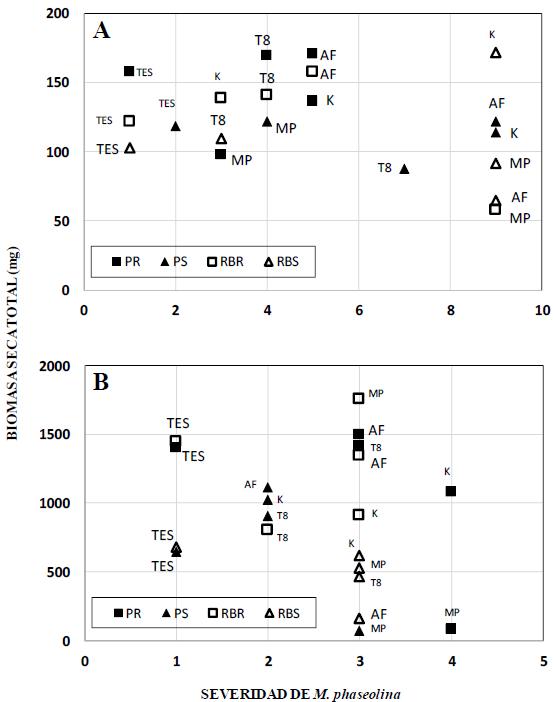
Figure 1. Relationship between the severity of charcoal rot and the total dry biomass accumulated in the sixth evaluation in sorghum grown in a greenhouse. (A) Experiment 1, (B) Experiment 2. PR=Pioneer 8282/Irrigation; PS=Pioneer 8282/Water deficit; RBR=RB Pattern/Irrigation; RBS=RB Pattern/Water.
  • Vol. 2025, 43(4)
  • Phytopathological Note
  • Special Issue

Trichoderma afroharzianum and homeopathic substances on the in vitro growth of fungi associated with fungal diseases

byMiguel Ángel Ruiz González, Miguel Ángel Serrato Cruz*, Roney Solano Vidal, Ernestina Valadez Moctezuma

Received: 19/November/2024 – Published: 25/September/2025
DOI: https://doi.org/10.18781/R.MEX.FIT.2024-11

Abstract Background/Objective. Trichoderma spp. is a sustainable alternative, as well as some homeopathic substances that are emerging as potential antifungal agents; making them promising solutions for combating and controlling diseases in crops of economic importance. The objective of the study was to evaluate the antagonism of Trichoderma afroharzianum and homeopathic substances against four fungi responsible for diseases of horticultural importance.

Materials and Methods. The antagonism of T. afroharzianum against Phytophthora capsici, Rhizoctonia solani, Botrytis cinerea, and Fusarium oxysporum was evaluated using the dual culture technique. The percentage of inhibition was estimated every 24 hours by measuring mycelial growth and type of antagonism. Homeopathic substances derived from essential oil of Tagetes remotiflora, Tagetes arenicola, and Tagetes lucida were evaluated using the poisoned agar technique on all organisms. A completely randomized design with four replicates was used. The data were analyzed using Tukey's mean comparison test (p ≤ 0.05).

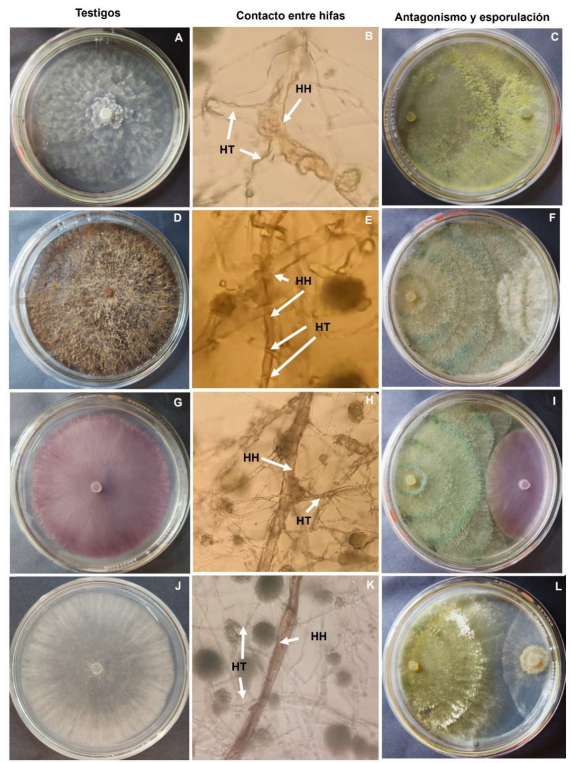
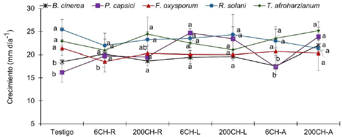
Results. T. afroharzianum inhibited over 50% of the fungi and stopped their growth in all cases. Only in the interaction between P. capsici and T. afroharzianum did the antagonist grow over P. capsici and deform the hyphae. Homeopathic oil of T. remotiflora in 6CH dinamization reduced the growth of R. solani (21.95 vs 25.38 mm in the control), F. oxysporum (18.52 vs 21.35 mm in the control), and T. afroharzianum (20.87 vs 22.86 mm in the control), while 200CH dinamization of T. arenicola stimulated the growth of T. afroharzianum (25.15 vs 22.86 mm in the control), P. capsici (23.85 vs 16.18 mm in the control) and B. cinerea (25.98 vs 18.48 mm in the control).

Conclusion. T. afroharzianum showed a greater ability to counteract the growth of P. capsici and B. cinerea. Homeopathic substances may have applications as inhibitors and stimulators of mycelial growth; further research is needed to better guide their applications

Show Figures and/or Tables
Figure 1. Mycelial growth inhibition of <em>B. cinerea</em> (5 days),<em> P. capsici</em> (11 days), <em>R. solani</em> (8 days), and <em>F. oxysporum</em> (9 days) due to the effect of <em>T. afroharzianum</em> under <em>in vitro</em> conditions. Mean ± standard deviation (n=4) with different letters are statistically different (Tukey, p ≤ 0.05).
Figure 1. Mycelial growth inhibition of B. cinerea (5 days), P. capsici (11 days), R. solani (8 days), and F. oxysporum (9 days) due to the effect of T. afroharzianum under in vitro conditions. Mean ± standard deviation (n=4) with different letters are statistically different (Tukey, p ≤ 0.05).
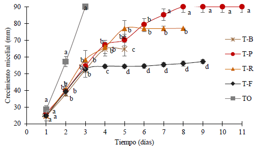
Figure 2. <em>In vitro</em> mycelial growth (mm) of <em>T. afroharzianum</em> in dual culture against <em>B. cinerea</em> (B), <em>R. solani</em> (R), <em>P. capsici</em> (P), and <em>F. oxysporum</em> (F). TO: control, T: <em>T. afroharzianum</em>, Mean ± standard deviation (n=4) with different letters are statistically different (Tukey, p ≤ 0.05).
Figure 2. In vitro mycelial growth (mm) of T. afroharzianum in dual culture against B. cinerea (B), R. solani (R), P. capsici (P), and F. oxysporum (F). TO: control, T: T. afroharzianum, Mean ± standard deviation (n=4) with different letters are statistically different (Tukey, p ≤ 0.05).
Figure 3. Individual mycelial growth (mm) of controls: <em>P. capsici</em> (A), <em>R. solani</em> (D), <em>F. oxysporum</em> (G) and <em>B. cinerea</em> (J) and antagonism in dual culture at the moment of hyphal contact (B, E, H, K) and sporulation after the controls covered the Petri dish (C, F, I, L). HH: fungal hyphae, HT: <em>T. afroharzianum</em> hyphae.
Figure 3. Individual mycelial growth (mm) of controls: P. capsici (A), R. solani (D), F. oxysporum (G) and B. cinerea (J) and antagonism in dual culture at the moment of hyphal contact (B, E, H, K) and sporulation after the controls covered the Petri dish (C, F, I, L). HH: fungal hyphae, HT: T. afroharzianum hyphae.
Figure 4. Fungal growth dynamics due to the effect of homeopathic substances on<em> B. cinerea</em>, <em>P. capsici</em>, <em>F. oxysporum</em>, <em>R. solani</em>, and <em>T. afroharzianum</em>. CH: centesimal hahnemannian, R: <em>T. remotiflora</em>, L: <em>T. lucida</em>, A: <em>T. arenicola</em>. Mean ± standard deviation (n=4) with different letters are statistically different (Tukey, p ≤ 0.05).
Figure 4. Fungal growth dynamics due to the effect of homeopathic substances on B. cinerea, P. capsici, F. oxysporum, R. solani, and T. afroharzianum. CH: centesimal hahnemannian, R: T. remotiflora, L: T. lucida, A: T. arenicola. Mean ± standard deviation (n=4) with different letters are statistically different (Tukey, p ≤ 0.05).
  • Vol. 2025, 43(4)
  • Scientific Article
  • Special Issue

Microbial control of Meloidogyne incognita on Capsicum chinense under an organic production system

byCitlally Guadalupe Puc Flores, Felicia Amalia Moo Koh, José M Tun Suárez, Eduardo Villanueva Couoh, Jairo Cristóbal Alejo*

Received: 23/November/2024 – Published: 25/September/2025
DOI: https://doi.org/10.18781/R.MEX.FIT.2024-33

Abstract Background/objective. Habanero pepper (Capsicum chinense) cultivation is limited by root-knot nematodes such as Meloidogyne incognita, and its control is achieved through the repeated application of synthetic nematicides. An alternative is the application of microbial agents. The objective of this study was to evaluate the control of M. incognita with native Trichoderma spp. in C. chinense, in an organic production system.

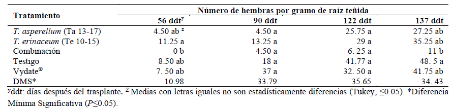
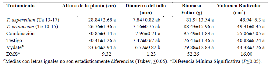
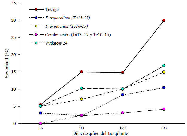
Materials and Methods. Seedlings of the 45-day-old Izamal cultivar were used and transplanted with a substrate based on soil, bocashi and volcanic rocks (6:3:1), three inoculations of native strains of Trichoderma asperellum (Ta13-17), T. erinaceum (Te10-15) and their combination were applied at the time of transplantation, and eight and 15 days later, as control treatments: a nematicide (Vydate®) and water. Four destructive samplings were carried out during the crop cycle, and the variables were the severity with the galling index, number of eggs and females per g of root.

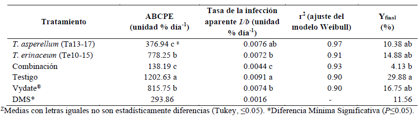
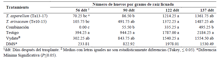
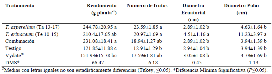
Results. In relation to the control, the combination of T. asperellum (Ta13-17) and T. erinaceum (Te10-15) caused significantly lower severity of the nematode estimated with the AUDPC. The apparent infection rate using the Weibull model (1/b) and final galling index (4.13%), also showed the lowest average reproduction of the nematode (number of eggs and females per g of root). With the combination of T. asperellum (Ta13-17) and T. erinaceum (Te10-15) the crop yield was significantly improved by 47.26 and 34.25 % in relation to the control and Vydate®, respectively.

Conclusion. In an organic production system of habanero pepper for the control of M. incognita; the individual application of T. asperellum (Ta13-17) significantly decreased the gall index; however, when combined with T. erinaceum (Te10-15) it improved nematode control (86.17%).

Show Figures and/or Tables
Table 1. Effect of treatments on the control of <em>M. incognita</em> estimated with epidemiological parameters in the organic cultivation of <em>C. chinense.</em>
Table 1. Effect of treatments on the control of M. incognita estimated with epidemiological parameters in the organic cultivation of C. chinense.
Table 2. Effect of treatments on the number of <em>M. incognita</em> eggs in organic cultivation of <em>C. chinense</em>.
Table 2. Effect of treatments on the number of M. incognita eggs in organic cultivation of C. chinense.
Table 3. Effect of treatments on the number of <em>M. incognita</em> females in organic cultivation of <em>C. chinense</em>.
Table 3. Effect of treatments on the number of M. incognita females in organic cultivation of C. chinense.
Table 4. Effect of treatments in the control of <em>M. incognita</em> on the agronomic variables of the organic cultivation of <em>C. chinense</em>.
Table 4. Effect of treatments in the control of M. incognita on the agronomic variables of the organic cultivation of C. chinense.
Table 5. Effect of treatments on fruit yield in <em>C. chinense</em> cultivation.
Table 5. Effect of treatments on fruit yield in C. chinense cultivation.
Figure 1. Curves of the progress of the gall index in the organic cultivation of <em>C. chinense</em>, during the period of 56, 90, 122 y 137 days after transplant.
Figure 1. Curves of the progress of the gall index in the organic cultivation of C. chinense, during the period of 56, 90, 122 y 137 days after transplant.
  • Vol. 2025, 43(4)
  • Scientific Article
  • Special Issue

Evaluation of the ΦXaF18 bacteriophage formulation for the control of Xanthomonas euvesicatoria in chili pepper plants under field conditions

byGabriel Ibarra Rivera, Evangelina Esmeralda Quiñones Aguilar, Guillermo Alejandro Solís Sánchez, Eristeo García Márquez, Gabriel Rincón Enríquez*

Received: 20/November/2024 – Published: 08/September/2025
DOI: https://doi.org/10.18781/R.MEX.FIT.2024-12

Abstract Background/Objective. Xanthomonas euvesicatoria (Xe) is the causal agent of bacterial spot in chili pepper crops, a disease that is commonly managed with agricultural antibiotics. As a biological alternative, bacterial viruses (bacteriophages) offer a promising approach to the control of plant pathogenic bacteria. The objective of this study was to evaluate the biological effectiveness of the bacteriophage ΦXaF18 formulated with UV radiation protection (Fagolytic) for the control of Xe in chili pepper plants under open field conditions and in commercial field plots.

Materials and Methods. Experiments were conducted in block or completely random arrangement; five treatments were evaluated under open-field and field conditions: 1) Fagolytic, 2) Unformulated ΦXaF18 bacteriophage, 3) LS formulation with ΦXaF18 bacteriophage, 4) Kasumin® agricultural antibiotic, 5) Untreated control. The number of leaves with symptoms, number of chlorotic spots and the severity index were measured with a 6-level ordinal scale (0-5) and viral concentration in foliage. Data were statistically analyzed with an analysis of variance and a Tukey test or a nonparametric Kruskal-Wallis test (p ≤ 0.05).

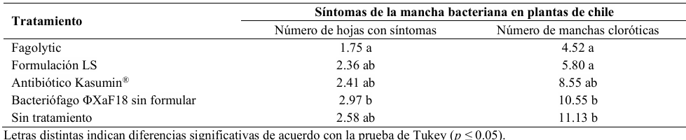
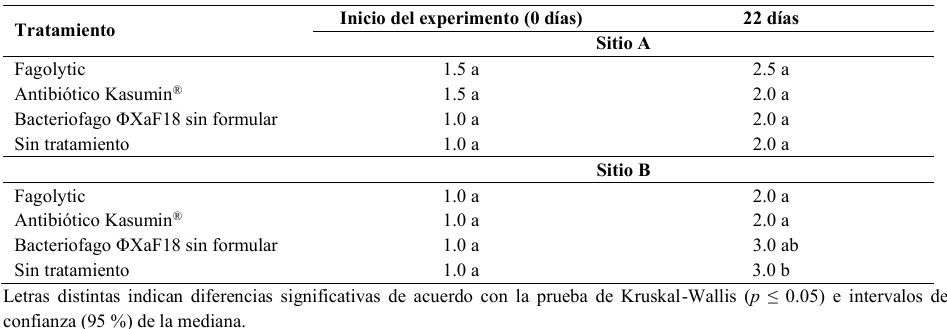
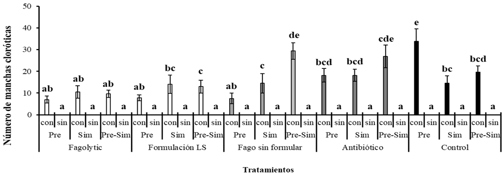
Results. Fagolytic reduced bacterial spot severity by 32% and chlorotic spots by 57% compared to unformulated bacteriophage ΦXaF18 under open-field conditions. In the field, Fagolytic significantly decreased the severity index in site B of experimental plot 1 (p ≤ 0.05), with an efficacy comparable to the antibiotic Kasumin®. In addition, at site A, Fagolytic showed a significantly higher viral concentration of ΦXaF18 on the foliage at 48 h after the application of the treatments. Whereas at the time of application (preventive, simultaneous or combined) it had no significant effect on the severity of the disease.

Conclusion. Fagolytic proved to be an effective biological alternative for the control of bacterial spots (caused by Xe) in chili pepper plants, by reducing the severity of the disease and maintaining its persistence in the foliage, with comparable results to the Kasumin® antibiotic both under open-field conditions and in commercial plots at the field level.

Show Figures and/or Tables
Table 1. Description of the ordinal severity scale used to evaluate bacterial spot (<em>X. euvesicatoria</em>) in chili pepper plants in commercial field plots located in Mascota, Jalisco.
Table 1. Description of the ordinal severity scale used to evaluate bacterial spot (X. euvesicatoria) in chili pepper plants in commercial field plots located in Mascota, Jalisco.
Table 2. Effect of biological treatments for the management of bacterial spot in chili pepper plants under open-field/greenhouse conditions.
Table 3. Effect of application timing of treatments for the management of bacterial spot in chili pepper plants under open-field conditions.
Table 4. Evaluation of bacterial spot severity in chili pepper plants at 0 and 22 days after treatment application for disease control in commercial experimental plot 1 located in Mascota, Jalisco.
Table 5. Effect of treatments for the biological management of bacterial spot in chili pepper plants in commercial experimental plot 1 located in Mascota, Jalisco.
Table 6. Determination of viral concentration on chili pepper foliage at sites A and B of commercial experimental plot 1, located in Mascota, Jalisco, 48 h after application of the ΦXaF18 bacteriophage treatment.
Table 7. Evaluation of bacterial spot severity in chili pepper plants at 0 and 22 days after treatment application in commercial experimental plot 2 located in Mascota, Jalisco.
Figure 1. Effect of different treatments on the management of bacterial spot in chili pepper plants under greenhouse and open field conditions. Different letters indicate significant differences according to Tukey’s test (p ≤ 0.05). Bars in each rectangle represent ± standard error. With Xe inoculum (con), without Xe inoculum (sin). Pre: Preventive. Sim: Simultaneous. Pre-Sim: Preventive and Simultaneous.
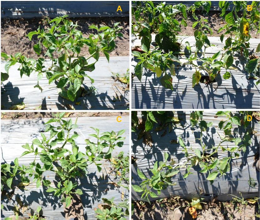
Figure 2. Visual comparison of bacterial spot severity in chili pepper plants treated with Fagolytic (A and C) and untreated control (B and D) at 22 days after the start of the experiment in commercial experimental plot 1 located in Mascota, Jalisco.
  • Vol. 2025, 43(3)
  • Phytopathological Note

Sensitivity of Sclerotium rolfsii, the causal agent of southern blight in chili pepper, to Trichoderma spp., plant extracts, and fungicides

byJosé Francisco Díaz Nájera, José Terrones Salgado*, Sergio Ayvar Serna, Mateo Vargas Hernández, Maricela Apáez Barrios, José Luis Arispe Vázquez, José Francisco Adame Bores

Received: 19/March/2025 – Published: 31/August/2025
DOI: https://doi.org/10.18781/R.MEX.FIT.2209-1

Abstract Background/Objective. Chili pepper (Capsicum annuum) are widely grown in Mexico. They are affected by facultative soil pathogens, among which the fungus Sclerotium rolfsii can be critical. The objectives of this research were to identify culturally, morphologically, and molecularly the causal agent of southern blight in Guerrero native chili pepper and to evaluate the in vitro antibiosis of Trichoderma spp., as well as the sensitivity of the causal agent to fungicides and plant extracts.

Materials and Methods. At CEP-CSAEGRO, plants of three types of local chili pepper genotypes with symptoms of southern blight were collected from which 20 fungal isolates were isolated, purified, and characterized. One isolate was selected for molecular identification. DNA was extracted using a commercial kit and amplified by PCR, and the internal transcribed spacer (ITS) region was subsequently sequenced. The sequences were edited, and a BLAST analysis and comparison with the NCBI database were performed. To confirm pathogenicity, Koch's postulates were performed in duplicate on chili seedlings of a local genotype. Using a completely randomized experimental design, the in vitro antibiotic activity of Trichoderma spp. against the causal agent was evaluated, as well as its in vitro sensitivity to fungicides and plant extracts.

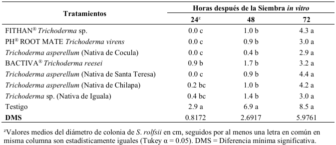
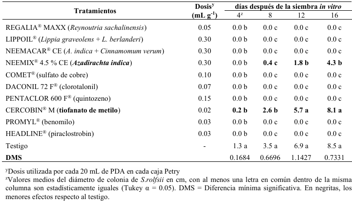
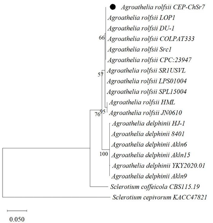
Results. Cultural, morphological, and molecular characterization (PV569903, NCBI accession number) identified Agroathelia rolfsii (S. rolfsii) as the causal agent of southern blight on native chili peppers. Pathogenicity tests showed symptoms identical to those identified in the field. In antibiosis trials, the highest mycelial inhibition of S. rolfsii was obtained with T. virens (65.18%), Trichoderma sp. (65.18%), and T. asperellum (66%). In the in vitro fungicide sensitivity, complete mycelial inhibition (100%) was obtained with copper sulfate, chlorothalonil, quintozene, benomyl, and pyraclostrobin. Commercial plant extracts from Reynoutria sachalinensis, Lippia graveolens + L. berlanderi, and A. indica + Cinnamomum verum completely inhibited the mycelial development of S. rolfsii (100%).

Conclusion. S. rolfsii is the pathogen that causes southern blight in native chili peppers in Guerrero. Trichoderma asperellum (native to Cocula), Trichoderma sp. (native to Iguala), and T. virens (PHC® ROOTMATE) inhibited the growth of S. rolfsii, as did some commercial fungicides and plant extracts. These are suggested as promising alternatives, subject to in vivo validation in the field, for the management of this fungal pathogen under a sustainable integrated approach.

Show Figures and/or Tables
Table 1. <em>In vitro</em> antibiotic activity - PDA with seven <em>Trichoderma</em> spp. products/strains against <em>S. rolfsii</em> isolated from local Guerrero chili peppers (<em>Capsicum annuum</em>).
Table 1. In vitro antibiotic activity - PDA with seven Trichoderma spp. products/strains against S. rolfsii isolated from local Guerrero chili peppers (Capsicum annuum).
Table 2. <em>In vitro</em> sensitivity – PDA of <em>Sclerotium rolfsii</em>, isolated from Guerrero native chili pepper (<em>Capsicum annuum</em>), to commercial fungicides and plant extracts evaluated at four 4-days intervals.
Table 2. In vitro sensitivity – PDA of Sclerotium rolfsii, isolated from Guerrero native chili pepper (Capsicum annuum), to commercial fungicides and plant extracts evaluated at four 4-days intervals.
Figure 1. Phylogenetic tree constructed for diagnostic purposes from sequences of the internal transcribed space (ITS) region of <em>Agroathelia</em> spp. isolates from the NCBI and the isolate found in Guerrero CEP-ChSr7 (in black circle). Maximum likelihood algorithm in MEGA 11, with Bootstrap a1000 simulations.
Figure 1. Phylogenetic tree constructed for diagnostic purposes from sequences of the internal transcribed space (ITS) region of Agroathelia spp. isolates from the NCBI and the isolate found in Guerrero CEP-ChSr7 (in black circle). Maximum likelihood algorithm in MEGA 11, with Bootstrap a1000 simulations.
  • Vol. 2025, 43(3)
  • Phytopathological Note

First report of dieback in jackfruit (Artocarpus heterophyllus) caused by Lasiodiplodia theobromae in Nayarit, Mexico

byAntonio Betancourt Aranguré, Gregorio Luna Esquivel*, Graciela Guadalupe López Guzmán, Claudio Rios Velasco, Octavio Jhonathan Cambero Campos, Elia Cruz Crespo, Eliana Andrea Martínez Mera

Received: 11/March/2024 – Published: 31/August/2025
DOI: https://doi.org/10.18781/R.MEX.FIT.2403-3

Abstract Background/Objective. Jackfruit (Artocarpus heterophyllus) is an economically important fruit for Nayarit, accounting for over 90% of the national crop. Branch dieback has been detected in jackfruit trees in recent years, so the objective of this study was to identify the causative agent of branch dieback in A. heterophyllus in the state of Nayarit, Mexico.

Materials and Methods. From a collection of Lasiodiplodia theobromae strains from commercial orchards of jackfruit, mango (Mangifera indica), soursop (Annona muricata), avocado (Persea americana), and Persian lime (Citrus latifolia) in Nayarit, the pathogenicity of L. theobromae was evaluated on one-year-old jackfruit trees. The isolates were molecularly identified by sequencing the internal transcribed spacer (ITS5 and ITS4 primers) and translation elongation factor 1-alpha (TEF1; primers EF-728/EF-986) regions.

Results. All isolates were pathogenic to jackfruit. LY1 and LG4, from jackfruit and soursop, respectively, showed high aggressiveness on branches. Dieback symptoms were evident two months post-inoculation, with shoot rot, internal necrotic lesions, and bark fissures on branches and stems, as well as tree death. The ITS and EF1-α sequences of isolates recovered from pathogenicity testing showed 99–100% similarity to previously published sequences for L. theobromae in GenBank.

Conclusion. This is the first report of L. theobromae as a causal agent of dieback of jackfruit A. heterophyllus in Nayarit, Mexico.

Show Figures and/or Tables
Table 1. Location and botanical source of <em>Lasiodiplodia theobromae</em> isolates used in pathogenicity tests to determine the etiological implication in branch dieback of jackfruit (<em>Artocarpus heterophyllus</em>).
Table 1. Location and botanical source of Lasiodiplodia theobromae isolates used in pathogenicity tests to determine the etiological implication in branch dieback of jackfruit (Artocarpus heterophyllus).
Figure 1. A. <em>Lasiodiplodia</em> culture on day 3 in PDA. B. <em>Lasiodiplodia</em> culture after 7 days in PDA. C. Septated maroon brown hyphae viewed under a compound microscope at 40X. D. Conidia, characteristically two-celled and dark, at maturity under a compound microscope at 40X.A. <em>Lasiodiplodia</em> culture on day 3 in PDA. B. <em>Lasiodiplodia</em> culture after 7 days in PDA. C. Septated maroon brown hyphae viewed under a compound microscope at 40X. D. Conidia, characteristically two-celled and dark, at maturity under a compound microscope at 40X.
Figure 1. A. Lasiodiplodia culture on day 3 in PDA. B. Lasiodiplodia culture after 7 days in PDA. C. Septated maroon brown hyphae viewed under a compound microscope at 40X. D. Conidia, characteristically two-celled and dark, at maturity under a compound microscope at 40X.A. Lasiodiplodia culture on day 3 in PDA. B. Lasiodiplodia culture after 7 days in PDA. C. Septated maroon brown hyphae viewed under a compound microscope at 40X. D. Conidia, characteristically two-celled and dark, at maturity under a compound microscope at 40X.
  • Vol. 2025, 43(3)
  • Phytopathological Note

Diversity of endophytic bacteria in strawberry plants (Fragaria sp.) in Sinaloa

byRosa M Longoria Espinoza*, Rubén Félix Gastélum, Dulce R Hernández Luna

Received: 09/August/2024 – Published: 30/August/2025
DOI: https://doi.org/10.18781/R.MEX.FIT.2408-2

Abstract Background/Objective. Currently, agricultural systems require management strategies based on crop sustainability. Plant growth-promoting bacteria are an environmentally friendly option to reduce the use of chemicals in agriculture and promote sustainable agricultural practices. The objective of this research was to isolate and identify endophytic bacteria in strawberry plants (Fragaria x ananassa) cv Chanler, grown under organic conditions and to evaluate in vitro their ability to produce metabolites related to plant growth promotion.

Materials and Methods. Twenty-five healthy-looking strawberry plants of the Chanler variety were collected during the fall-winter 2023-2024 cycle. Genomic DNA was isolated from samples composed of root, stem, and leaf tissue, and the 16S rDNA gene was amplified.

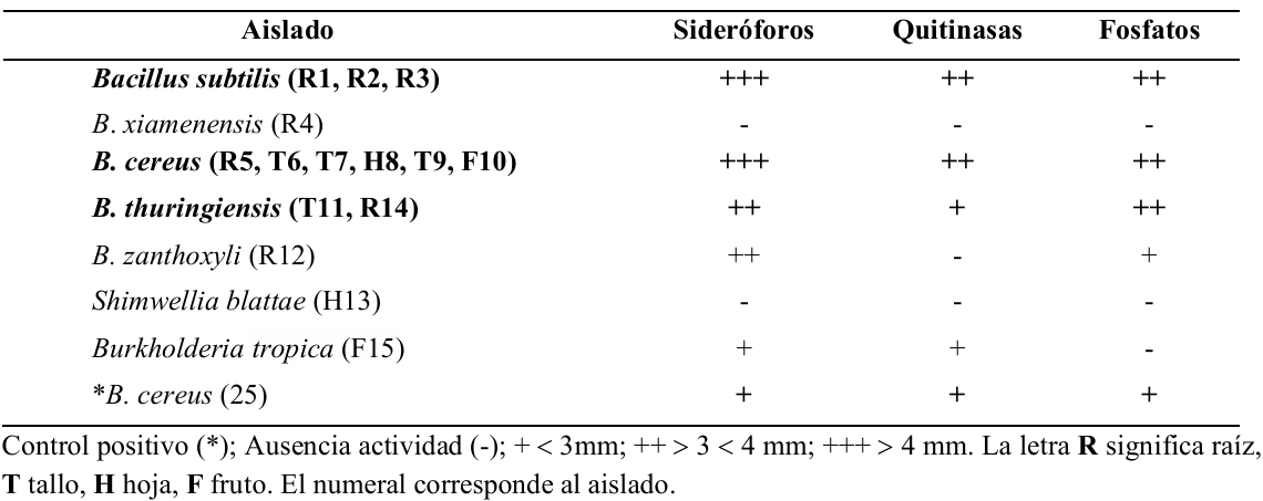
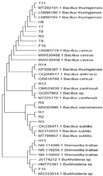
Results. Analysis of genomic sequences identified bacterial genera associated with the phylum Firmicutes, with the genus Bacillus predominating, along with the species B. cereus; B. subtilis, B. thuringiensis, B. xiamenensis, and B. zanthoxyli. Shimwellia blattae and Burkholderia tropica were detected in smaller numbers. Of these isolates, B. cereus, B. subtilis, and B. thuringiensis, in that order, showed the ability to produce siderophores, chitinase, and solubilize phosphorus in vitro.

Conclusion. B. cereus, B. subtilis, and B. thuringiensis exhibited metabolic characteristics related to plant growth promotion in vitro. These results in the existence of a diversity of endophytic bacteria in commercial strawberries. This provides information for understanding the endophytic bacteria-plant interaction in the specific environment of strawberry cultivation in the northern state of Sinaloa.

Show Figures and/or Tables
Table 1. Qualitative <em>in vitro</em> siderophore, chitinase, and phosphate production activity of six species of endophytic bacteria isolated from Chanler strawberry plants grown under organic production. Municipality of Ahome, Sinaloa, Mexico.
Table 1. Qualitative in vitro siderophore, chitinase, and phosphate production activity of six species of endophytic bacteria isolated from Chanler strawberry plants grown under organic production. Municipality of Ahome, Sinaloa, Mexico.
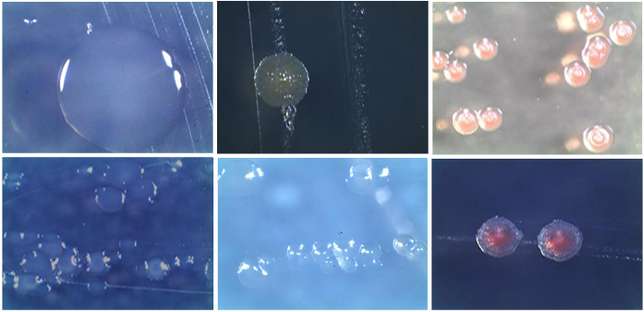
Figure 1. <em>In vitro</em> colonial morphology of endophytic bacteria isolated from the roots, stems, leaves, and fruit of strawberry plants (<em>Fragaria</em> x <em>ananassa</em>) cv. Chanler, viewed a digital stereoscope after 48 h of growth on nutrient agar. Note: The different morphology: smooth, translucent edges, circular shape, shiny surface, and coloration.
Figure 1. In vitro colonial morphology of endophytic bacteria isolated from the roots, stems, leaves, and fruit of strawberry plants (Fragaria x ananassa) cv. Chanler, viewed a digital stereoscope after 48 h of growth on nutrient agar. Note: The different morphology: smooth, translucent edges, circular shape, shiny surface, and coloration.
Figure 2. Dendrogram obtained using the <em>Neighbor</em>-<em>Joining</em> algorithm from sequences of the gene encoding the 16S rDNA subunit obtained from endophytic bacteria isolated from strawberry plants (<em>Fragaria</em> x <em>ananassa</em>), cv. Chanler, from organic commercial crops. Municipality of Ahome, Sinaloa, Mexico. The isolates obtained in this research are identified with acronyms and numbers: R-n: root; T-n: stem; H-n: leaf; F-n: fruit.
Figure 2. Dendrogram obtained using the Neighbor-Joining algorithm from sequences of the gene encoding the 16S rDNA subunit obtained from endophytic bacteria isolated from strawberry plants (Fragaria x ananassa), cv. Chanler, from organic commercial crops. Municipality of Ahome, Sinaloa, Mexico. The isolates obtained in this research are identified with acronyms and numbers: R-n: root; T-n: stem; H-n: leaf; F-n: fruit.
  • Vol. 2025, 43(3)
  • Scientific Article

Characterization of Sclerotium rolfsii and species of Trichoderma in commercial crops of potato in Sonora and Sinaloa, Mexico

byMaría Belén Irazoqui Acosta, Rubén Felix Gastelum*, Karla Yeriana Leyva Madrigal, Rosa Maria Longoria Espinoza, Gabriel Herrera Rodríguez, Sara Elodia Armenta López

Received: 09/August/2024 – Published: 28/August/2025
DOI: https://doi.org/10.18781/R.MEX.FIT.2408-1

Abstract Background/Objective. Sclerotium rolfsii causes losses in potato production up to 20% in the states of Sonora and Sinaloa. No studies exist supported by the scientific method on the identification of the fungus in the crop, neither of the species of Trichoderma present in the soil with potential to control the disease under field conditions. The objectives of the present study were to: a) obtain isolates of S. rolfsii from soil subject to the potato crop in the states of Sonora and Sinaloa, and determine the population density of sclerotia of the fungus; isolate and quantify the population density of Trichoderma spp., in the same soil to use them in subsequent studies for controlling the disease and determine the pathogenicity of S. rolfsii in potato tubers under laboratory conditions.

Materials and Methods. The isolates were collected from 239 commercial potato fields in the states of Sonora and Sinaloa. The population density was determined by counting the sclerotia of S. rolfsii and the colony forming units (CFU g-1) of Trichoderma in soil. The identification of the fungi was performed by morphological studies and molecular techniques including the internal transcribed spacer (ITS) for S. rolfsii and the subunit of RNA polymerase II (RPB2) for Trichoderma spp. The pathogenicity tests of the isolates of S. rolfsii were determined in potato tubers var. Fianna in the laboratory.

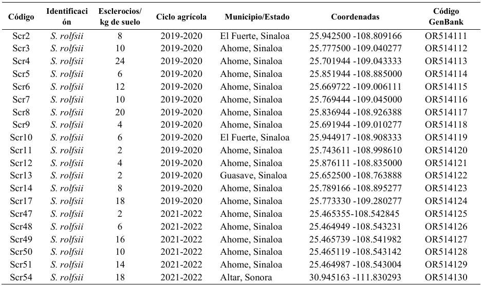
Results. Twenty isolates of S. rolfsii were identified whose population density varied from 2 to 24 sclerotia kg-1 of soil. On the other hand, 26 isolates of Trichoderma spp. were identified; 16 of them corresponded to T. asperellum, five to T. asperelloides, four to T. afroharzianum and one to T. azevedoi with a population density from 2 to 8 CFU g-1 of soil. The isolates of S. rolfsii resulted pathogenic to potato tubers var. Fianna, with different levels of aggressiveness.

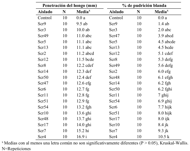
Conclusion. The results obtained in the present study indicate the presence of S. rolfsii in 8.4% of the 239 potato growing fields included in the study sampled in the growing cycles 2019-2022 con population densities from 2 to 24 sclerotia g-1 of soil. The pathogen confirmation by conventional and molecular techniques, as well as its capability to induce soft rot of potato tubers var. Fianna, provides evidence of its potential impact on potato production. Variable aggressiveness was detected among isolates, highlighting the Scr4 isolate as the most aggressive with penetration up to 16.9 mm and soft rotting of 10.5%. In parallel, 26 isolates of Trichoderma spp. were identified in 10.9% of the fields, with population densities from 2 to 8 CFU g-1 of soil. These findings highlight the importance of incorporating sustainable strategies for disease management, considering both the variability of the pathogen and the possible application of native antagonists; also, it is stressed the necessity to investigate the population dynamics of the pathogen and agricultural practices to minimize its survival.

Show Figures and/or Tables
Table 1. Identification, population density and origin of 20 <em>S. rolfsii</em> isolates obtained from soils in which potato is grown in Sonora and Sinaloa.
Table 1. Identification, population density and origin of 20 S. rolfsii isolates obtained from soils in which potato is grown in Sonora and Sinaloa.
Table 2. Identification ID, population density (CFU) and origin of 26 <em>Trichoderma</em> isolates sampled from soil of potato (<em>S. tuberosum</em>) commercial fields in Sonora and Sinaloa.
Table 2. Identification ID, population density (CFU) and origin of 26 Trichoderma isolates sampled from soil of potato (S. tuberosum) commercial fields in Sonora and Sinaloa.
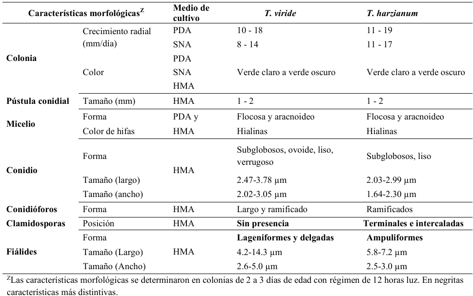
Table 3. Morphological characteristics of isolates of the <em>Trichoderma viride</em> and <em>Trichoderma harzianum</em> species complexes in three culture media.
Table 3. Morphological characteristics of isolates of the Trichoderma viride and Trichoderma harzianum species complexes in three culture media.
Table 4. Penetration of the pathogen and percentage of soft rot in potato tubers var. Fianna with artificial inoculation.
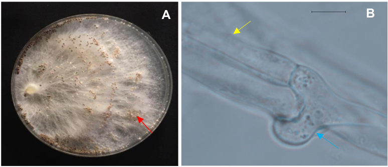
Figure 1. Morphological characteristics of <em>Sclerotium rolfsii</em> (Scr17). (A) Colony morphology in PDA medium, and sclerotia (red arrow); (B) Hyphal hyalin (yellow arrow) and fíbulae (blue arrow) (100x). Scale: (B= 50 µm).
Figure 1. Morphological characteristics of Sclerotium rolfsii (Scr17). (A) Colony morphology in PDA medium, and sclerotia (red arrow); (B) Hyphal hyalin (yellow arrow) and fíbulae (blue arrow) (100x). Scale: (B= 50 µm).
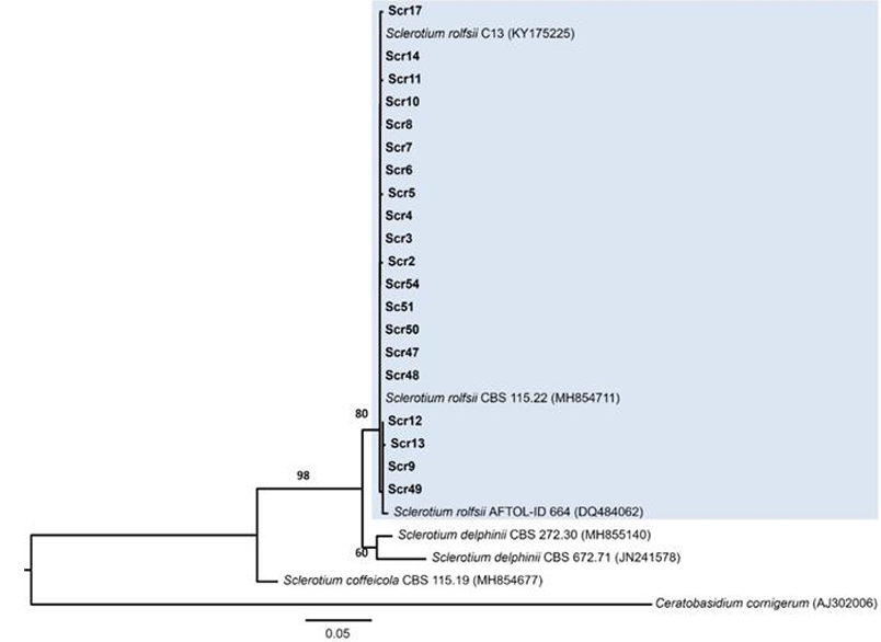
Figure 2. Phylogenetic analysis of <em>Sclerotium rolfsii</em> from Sonora and Sinaloa. Maximum likelihood tree based on the Internal Transcribed Spacer (ITS) of 20 <em>S. rolfsii</em> isolates. The isolates characterized in this study appear in bold letters. The bootstrap values over 50 are shown as percentages. The scale bar indicates the expected number of nucleotide substitutions.
Figure 2. Phylogenetic analysis of Sclerotium rolfsii from Sonora and Sinaloa. Maximum likelihood tree based on the Internal Transcribed Spacer (ITS) of 20 S. rolfsii isolates. The isolates characterized in this study appear in bold letters. The bootstrap values over 50 are shown as percentages. The scale bar indicates the expected number of nucleotide substitutions.
Figure 3. Colony and microscopic morphology of <em>Trichoderma asperellum</em> (TES19). (A) colony in PDA culture medium. (B) colony in SNA culture medium. (C) conidial pustules. (D and E) conidiophores and (F) conidia (100x). (C-F) in HMA medium. Scale: (C= 2 mm) (D= 10 µm) (E and F= 5 µm). This species belongs to <em>T. viride</em> complex.
Figure 3. Colony and microscopic morphology of Trichoderma asperellum (TES19). (A) colony in PDA culture medium. (B) colony in SNA culture medium. (C) conidial pustules. (D and E) conidiophores and (F) conidia (100x). (C-F) in HMA medium. Scale: (C= 2 mm) (D= 10 µm) (E and F= 5 µm). This species belongs to T. viride complex.
Figure 4. Colony and microscopic morphology of <em>Trichoderma afroharzianum</em> (TAF33). (A) colony in PDA culture medium. (B) colony in SNA culture medium. (C) conidial pustules. (D and E) chlamidospores and (F and G) conidiophores (H) conidia (100x). (C-H) in HMA medium. Scale: (C= 2 mm) (D and H= 5 µm) (E, F and G= 10 µm). This species belongs to <em>T. harzianum</em> complex.
Figure 4. Colony and microscopic morphology of Trichoderma afroharzianum (TAF33). (A) colony in PDA culture medium. (B) colony in SNA culture medium. (C) conidial pustules. (D and E) chlamidospores and (F and G) conidiophores (H) conidia (100x). (C-H) in HMA medium. Scale: (C= 2 mm) (D and H= 5 µm) (E, F and G= 10 µm). This species belongs to T. harzianum complex.
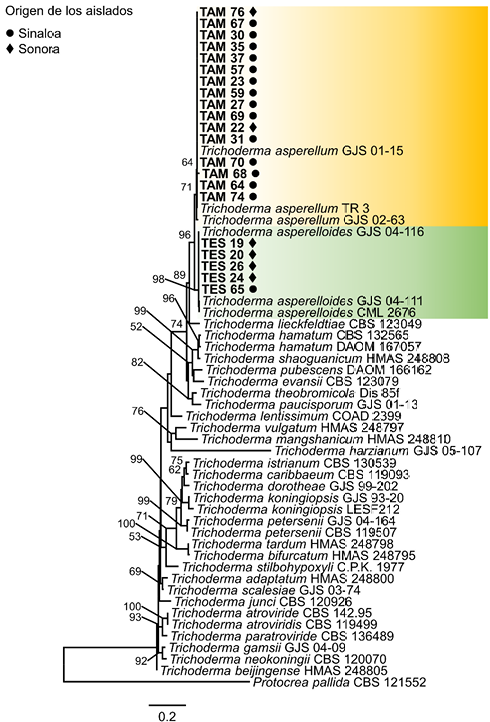
Figure 5. Maximum Likelihood phylogram of the <em>Trichoderma viride</em> species complex, based on the RNA polymerase II subunit (RPB2). The 21 <em>Trichoderma</em> isolates, obtained from potato plantations in Sonora and Sinaloa characterized in this study, appear in bold and highlighted in color. The bootstrap values >50 and the posterior probability values >0.5 are shown in the nodes. The scale bar indicates the expected number of nucleotide substitutions.
Figure 5. Maximum Likelihood phylogram of the Trichoderma viride species complex, based on the RNA polymerase II subunit (RPB2). The 21 Trichoderma isolates, obtained from potato plantations in Sonora and Sinaloa characterized in this study, appear in bold and highlighted in color. The bootstrap values >50 and the posterior probability values >0.5 are shown in the nodes. The scale bar indicates the expected number of nucleotide substitutions.
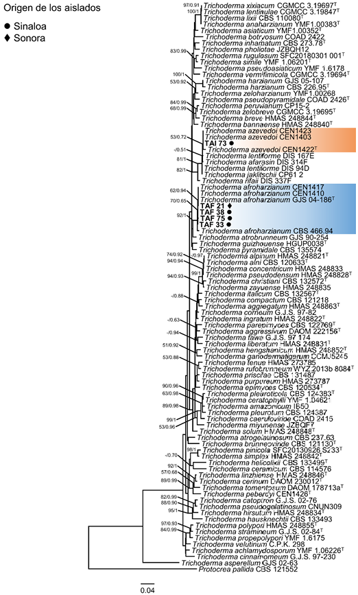
Figure 6. Maximum Likelihood phylogram of the <em>Trichoderma harzianum</em> species complex, based on the RNA polymerase II subunit (RPB2). The five <em>Trichoderma</em> isolates obtained from potato plantations in Sonora and Sinaloa characterized in this study, appear in bold and highlighted in color. The bootstrap values >50 and the posterior probability values >0.5 are shown in the nodes. The scale bar indicates the expected number of nucleotide substitutions.
Figure 6. Maximum Likelihood phylogram of the Trichoderma harzianum species complex, based on the RNA polymerase II subunit (RPB2). The five Trichoderma isolates obtained from potato plantations in Sonora and Sinaloa characterized in this study, appear in bold and highlighted in color. The bootstrap values >50 and the posterior probability values >0.5 are shown in the nodes. The scale bar indicates the expected number of nucleotide substitutions.
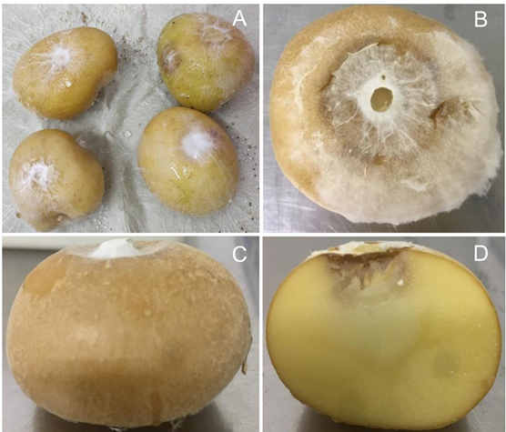
Figure 7. Symptoms of soft rot in Fianna variety potato tubers, caused by the isolate Scr17 of <em>S. rolfsii</em> by artificial inoculation. (A) Tubers with symptoms of <em>S. rolfsii</em> in a moist chamber. (B and C) lesions with cottonlike mycelium caused by the fungus. (D) Transverse cut of potato tuber with soft rot.
Figure 7. Symptoms of soft rot in Fianna variety potato tubers, caused by the isolate Scr17 of S. rolfsii by artificial inoculation. (A) Tubers with symptoms of S. rolfsii in a moist chamber. (B and C) lesions with cottonlike mycelium caused by the fungus. (D) Transverse cut of potato tuber with soft rot.

Page 5 of 16