Share this link via
Or copy link



Article filters
Search Papers:
byDarío Franco Romo, Norma Angélica Chávez Vela, José de Jesús Luna Ruiz*, Sylvia Patricia Fernández Pavía, José Francisco Morales Domínguez, Susana de la Torre Zavala
Received: 20/November/2025 – Published: 25/February/2026
DOI: https://doi.org/10.18781/R.MEX.FIT.2511-2
Abstract Background/Objective. Phytophthora capsici represents one of the most significant threats to pepper production worldwide, and its control relies predominantly on fungicide applications, which entail considerable environmental concerns. Therefore, exploring biological control strategies has become a relevant alternative. This study aimed to assess the potential of a bacterial isolate as a biocontrol agent against six P. capsici and one P. cinnamomi isolates.
Materials and Methods. Biocontrol effects were assessed in vitro using dual-culture assays between the bacterial isolate JJR198 and Phytophthora isolates to evaluate inhibition of mycelial growth, sporangia formation, and zoospore release, with additional observations by scanning electron microscopy. The efficacy of JJR198 in suppressing Phytophthora root rot was evaluated in pepper plants under greenhouse conditions by measuring disease severity in inoculated plants. In addition, salt tolerance and persistence of the bacterium in the rhizosphere and rhizoplane were determined. All data were subjected to ANOVA followed by Tukey’s test (p ≤ 0.05).
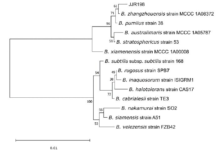
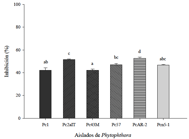
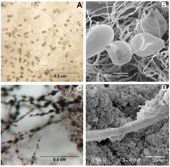
Results. Identified as Bacillus zhangzhouensis through 16S rRNA sequencing, the bacterial isolate JJR198, exhibited antagonistic activity. In vitro assays showed that strain JJR198 reduced mycelial growth by 42.0 to 52.7% across six tested Phytophthora isolates. The biocontrol activity depended on the initial bacterial concentration, as sporangia formation and zoospore release in the tested Phytophthora capsici isolates were completely suppressed at 1.0 × 10⁸ CFU mL-1, whereas no effect was observed at lower concentrations (1.0 × 10⁴ and 1.0 × 10⁶ CFU mL-1). Using optical and electron microscopy, it was observed that the hyphae and mycelia were covered by bacterial cells. However, in the in vivo evaluation, pretreatment of pepper plants with JJR198 (1.0 × 10⁸ CFU mL-1) at 7, 3, or 0 days before oomycete inoculation did not reduce disease severity caused by Phytophthora capsici. JJR198 also showed the ability to grow under high-salinity conditions, as it developed on NA medium containing 7.5% NaCl after 48 h of incubation at 30 °C. Additionally, the presence of the bacterial inoculum was confirmed in pepper roots: colonies with morphological characteristics identical to JJR198 were recovered from rhizosphere and rhizoplane samples, with counts of 1.6 × 10⁶ and 0.19 × 10⁶ CFU mL-1, respectively.
Conclusion. The results indicate that Bacillus zhangzhouensis JJR198 exhibits in vitro antagonistic activity against multiple Phytophthora isolates, achieving mycelial growth inhibition ranging from 42.0 to 52.7%, as well as inhibition of sporangia formation and zoospore release at a concentration of 1.0 × 10⁸ CFU mL⁻¹. However, its effectiveness in planta remained limited under the conditions tested.
Share this link via
Or copy link


byOlga Gómez Rodríguez, Edgar Villar Luna, Laith Khalil Tawfeeq Al-Ani, Iván Morales Soto, Reyna Vargas Abasolo, Liliana Aguilar Marcelino*
Received: 28/July/2025 – Published: 09/February/2026
DOI: https://doi.org/10.18781/R.MEX.FIT.2407-4
Abstract Background/Objective. Nematophagous mites represent a potentially effective tool in integrated management programs for plant-parasitic nematodes. The objective of this study was to evaluate the predatory capacity of the nematophagous mite Sancassania mycophaga against the plant-parasitic nematode Nacobbus aberrans under laboratory conditions.
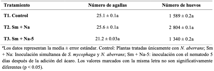
Experimental development. Two independent experiments were conducted. For this purpose, the experimental unit consisted of a pot with a pepper plant (Capsicum annuum), in which 500 or 1000 J2 juveniles of N. aberrans were inoculated. Four treatments were evaluated: (1) control (nematodes only), (2) simultaneous inoculation of mites and nematodes, (3) inoculation of nematodes five days after mites, and (4) inoculation of nematodes 10 days after mites. In all treatments, five mites were introduced per pot. The variables were: percentage of J2 penetration in roots, number of galls, eggs, and abundance of mites. The data were subjected to ANOVA and comparison of means (LSD, p < 0.05).
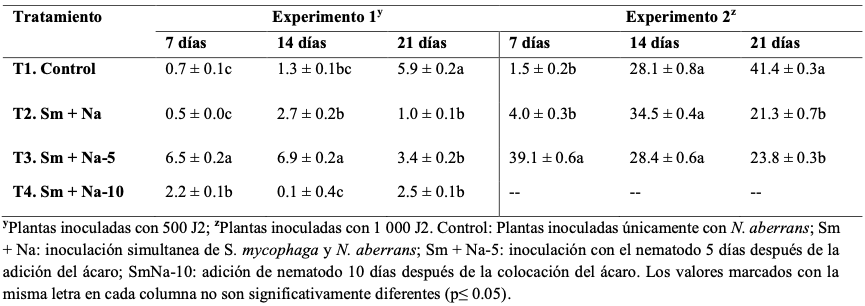
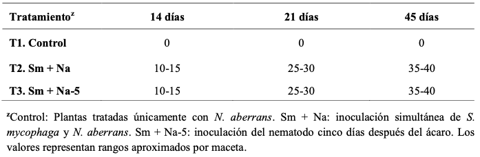
Results. Twenty-one days after the establishment of the trials, a significant reduction in nematode penetration was observed in all treatments with mites, compared to the control. In the experiment with 500 J2, the percentages of root penetration were 1.0, 3.4, and 2.5% for treatments 2, 3, and 4, respectively. In the experiment with 1000 J2, the penetration percentage was 21.3 and 23.8% for treatments 2 and 3. From day 14 onwards, the mite population showed sustained growth, increasing from an initial five individuals to more than 25 at 21 days (>400%) and more than 35 at 45 days (>600%). No significant differences in the number of galls or eggs were detected at 45 days.
Conclusions. S. mycophaga reduced the penetration of N. aberrans into pepper roots. With 500 J2, the relative reduction ranged from 42 to 83%, while with 1 000 J2 it varied between 42 and 49% compared to the control. In addition, the mite managed to establish itself and multiply in the experimental system. These results support its potential as a biocontrol agent for N. aberrans.
Share this link via
Or copy link


byLuis Alfonso Jiménez Ortega, Octavio Valdez Baro, Laura A Contreras Angulo, Erick P Gutiérrez Grijalva, Karina Valencia Sandoval, Raymundo Saúl García Estrada, José Basilio Heredia*
Received: 22/May/2025 – Published: 09/February/2026
DOI: https://doi.org/10.18781/R.MEX.FIT.2206-3
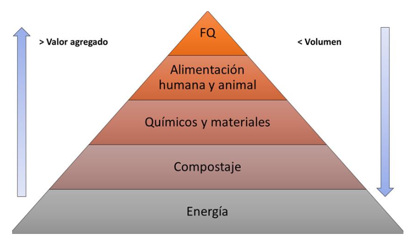
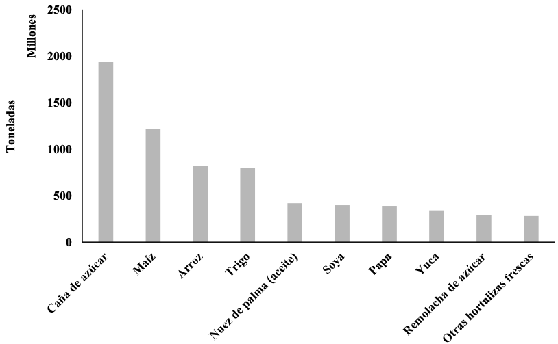
Abstract Justification. The use of chemical pesticides has been widespread for several decades, proving effective in controlling pests that reduce crop quality and productivity. However, due to their chemical nature, they present side effects that affect the health of the applicator, the consumer, and the environment. Therefore, the use of natural products is currently emerging as an alternative for controlling phytopathogenic diseases and pests. The objective of this research was to review and analyze the bactericidal and fungicidal effects of phytochemical-rich extracts obtained from agricultural and agro-industrial byproducts and waste.
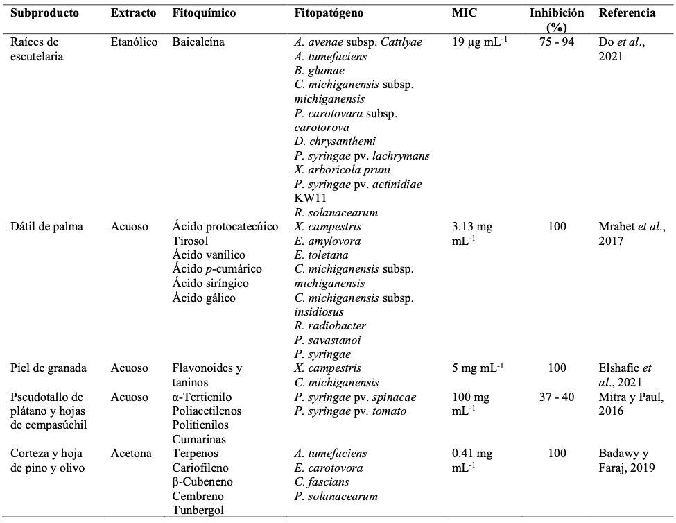
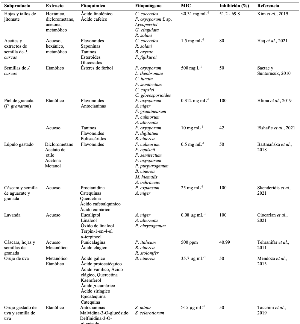
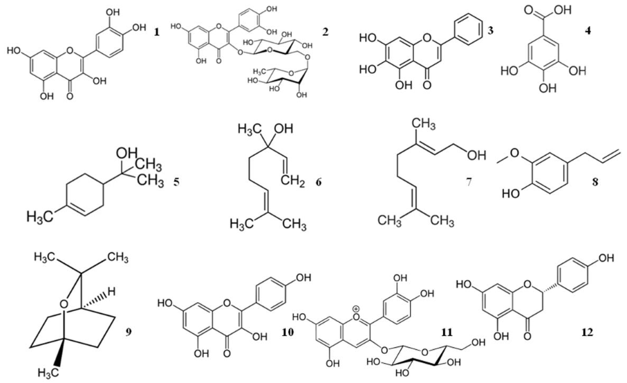
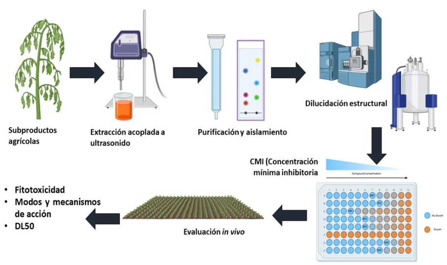
Theoretical and experimental framework. A literature review was conducted using Scopus and Web of Science, focusing on English- and Spanish-language publications from the last 10 years. Only publications related to the extraction of phytochemicals from agricultural and/or agro-industrial byproducts and that investigated their effect against phytopathogenic organisms were considered. In this regard, the main phytochemicals with antibacterial and antifungal activity are phenolic compounds such as flavonoids and other derivatives. Likewise, hydrophobic compounds such as terpenes (essential oils) have demonstrated biocidal potential at concentrations similar to those currently marketed. The main modes and mechanisms of action are multi-target, meaning they affect more than one metabolic pathway and organelle. For example, they affect the cell membrane by inserting into the lipid bilayer, reduce biofilm formation, reduce ergosterol synthesis, and attenuate chitin synthesis in the cell wall.
Conclusions and Perspectives. Phytochemicals with potent bactericidal and fungicidal activity were identified, primarily phenolic compounds and essential oils (terpenes). These phytochemicals exhibited IC50 values and reduction percentages even higher than those of the chemical controls. Agricultural byproducts and biomass, which are undervalued resources, represent an important alternative for extracting value-added phytochemicals that can be used across multiple industries, including agriculture, to develop sustainable, non-toxic biopesticides.
Share this link via
Or copy link

byGuadalupe Reyes García, Santo Ángel Ortega Acosta, Francisco Palemón Alberto*, Juan Elías Sabino López, Oscar Martín Antunez Ocampo, Claudia Martínez Alonso, Anayeli Morales Rosales
Received: 01/December/2025 – Published: 05/February/2026
DOI: https://doi.org/10.18781/R.MEX.FIT.2512-1
Abstract Background/Objective. Citrus (Citrus spp.) production is affected annually by anthracnose, associated with Colletotrichum spp. This disease can occur preharvest and postharvest and is generally controlled with synthetic fungicides, which pose health problems for farmers and consumers, negatively impact the environment, and lead to the emergence of resistant phytopathogen populations. An alternative to these problems is the application of plant extracts with antifungal properties, which could contribute to the production of safe food. Therefore, the objective of this study was to evaluate the effect of plant extracts under in vitro conditions against Colletotrichum gloeosporioides, causal agent of anthracnose on grapefruit (Citrus paradisi).
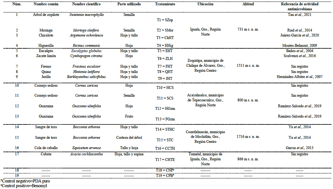
Materials and Methods. Extracts were obtained from 17 plant species. The effectiveness of the extracts was evaluated using the poisoned PDA culture technique (PDA + 10% plant extract), in which 5 mm diameter discs of the fungus were inoculated onto Petri dishes. The fungicide Benomyl (10%) was used as a positive control, and pure PDA as a negative control. A completely randomized experimental design with 19 treatments and three replicates was used. The percentage of inhibition of C. gloeosporioides mycelial growth was recorded when the negative control completely covered the surface of the Petri dish. Data were analyzed using the statistical software SAS version 9.0.
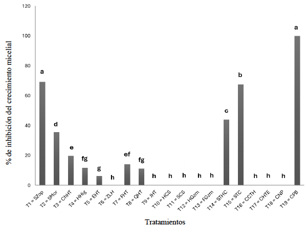
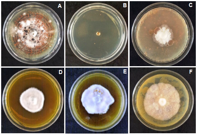
Results. The best extracts were zopilote seed (Swietenia macrophylla), bull blood tree bark (Bocconia arborea), and bull blood tree leaf and stem (B. arborea), and moringa seed (Moringa oleifera), inhibiting the growth of C. gloeosporioides by 69.2, 67.5, 44.1, and 35.6%, respectively. The positive control, Benomyl, inhibited C. gloeosporioides growth by 100%.
Conclusion: The best in vitro inhibitory effect on Colletotrichum gloeosporioides was obtained with Swietenia macrophylla seed extracts (69.2%), followed by Bocconia arborea bark (67.5%), B. arborea leaf and stem (44.1%), and Moringa oleifera (35.6%).
Share this link via
Or copy link
byCitlalmina Erandy Montes Hernández, Gustavo Mora Aguilera*, María Elizabeth Morales Cortés, Gerardo Acevedo Sánchez, Ángel López Zárate
Received: 14/June/2024 – Published: 31/December/2025
DOI: https://doi.org/10.18781/R.MEX.FIT.2406-5
Abstract Background/Objective. In strawberry (Fragaria x ananassa), the main commercial rosaceous fruit in Mexico, the occurrence of Neopestalotiopsis rosae (Ascomycota: Sporocadaceae) was recently reported in Michoacán. The objective was to analyze, using an etiological-epidemiological approach, the implication of two N. rosae ITS-haplotypes, associated to five isolates from strawberry and blackberry (Rubus sp.) selected for regional prevalence, in the pathogenesis, aggressivity, and inoculum load in strawberry cv. San Andreas.
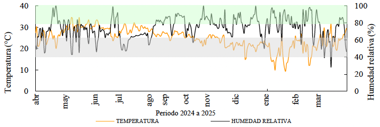
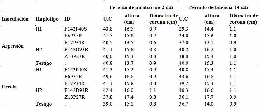
Materials and Methods. Three isolates of Haplotype 1 / Strawberry (H1): F142P40S, F6P35R, F17P34R, and two of Haplotype 2 (H2) / Blackberry: F142D93R and Z13P27R were cultured in V8 / 25° / 14 d until conidiomata formation, and in PDA / 25° / 7 d to obtain liquid and solid inoculum, respectively. Eight three-month-old strawberry plants cv. San Andreas were evaluated per isolate; two trifoliate leaves / 4 plants were inoculated with 4 mL spray 1 x 10⁶ conidia mL-1, and 4 plants with 5 x 5 mm mycelium PDA-blocks per crown wound. Sterile water was sprayed on 8 control plants. Plants were kept in a semi-covered greenhouse. Starting at 72 hai, every 3 d / 3 months, the following variables were measured: Number of lesions (LN), Lesion diameter (LD), Number of sporulating lesions (EL), Plant height, Crown diameter, and Chlorophyll units SPAD 502 (CU). The incubation period (Pi), latency period (Pl), and generation period (Pg) were estimated for each haplotype. Temperature and RH were recorded every 30 min with HOBO U23. Pathogenicity was validated using Koch's postulates. Isolates H1 and H2 were confirmed culturally by ITS1 / ITS4. ANOVA, in split plots design, and Tukey (p = 0.05) were conducted in RStudio.
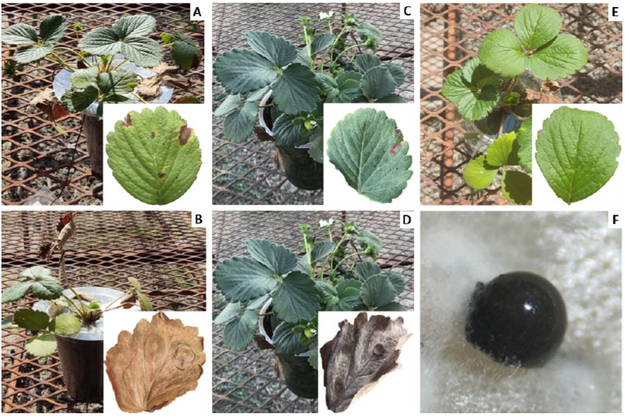
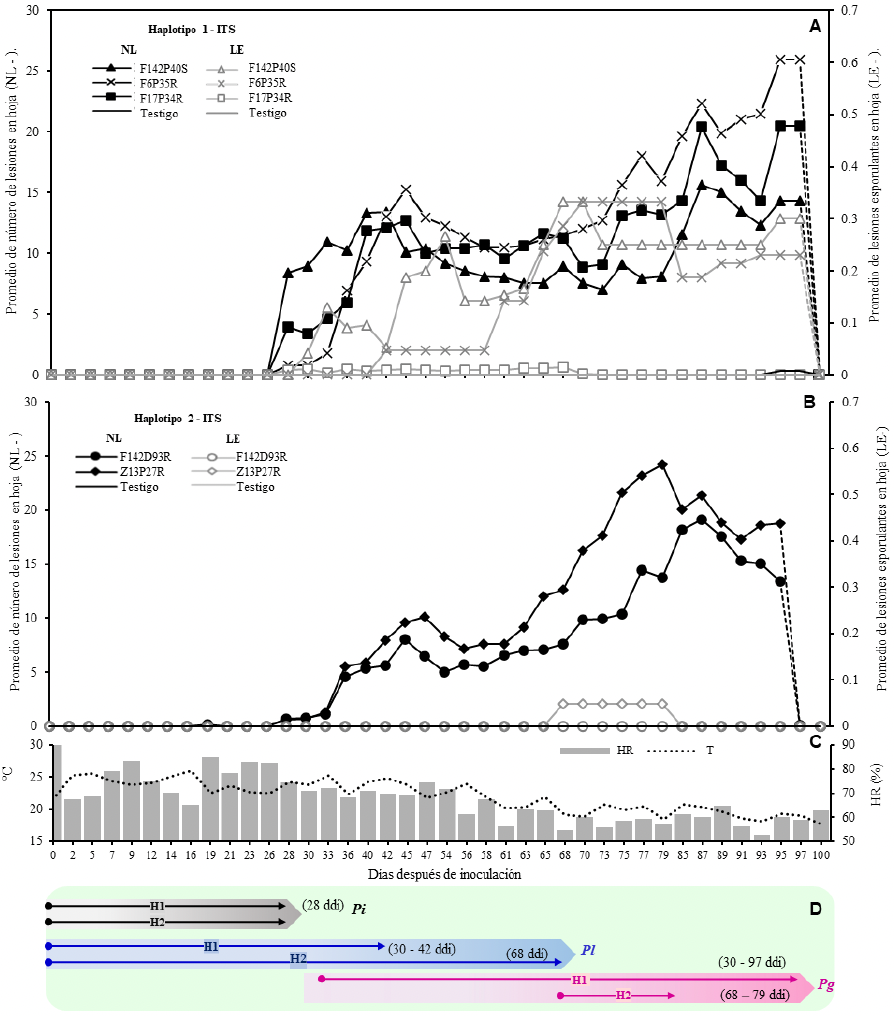
Results. The five isolates of N. rosae were more infectious on leaves (p = 0.0001) but exhibited variable and significant aggressivity (p = 0.0001) on LN, EL, and LD, with expression of circular leaf lesions, light brown color, dark edge, and reddish halo on the upper and lower surface. H1 isolates were more pathogenic than H2 (e.g., 3504 vs. 270 LN / plant), with H1 - F142P40S being more aggressive (p = 0.05), with an average LN = 187.5 and EL = 8.45 at 2 and 26 days after inoculation (dai), respectively. In contrast, H2 - F142D93R and H2 – Z13P27R had LN = 20 and 25 at 87 and 95 dai, respectively, with a maximum average range of 0.19 - 0.37 EL > 73 dai. Similarly, in crown, all isolates were pathogenic, but with less aggressivity and no statistical difference. A reddish-dark discoloration and internal necrotic rot was observed in crown and low root density. H1 - F17P34R was conspicuous with a maximum NL = 20 and LE < 0.1 at 85 dai. In leaf, the speed of symptoms expression and sporulation rate, although variable, was higher in isolates H1 vs. H2, determined, respectively, by 2 vs. 4 dai Pi, 14 - 16 (3 d) vs. 33 - 40 (7 d) dai Pl, and 14 - 73 (59 d) vs. 33 - 56 (23 d) dai Pg. The crown infection was slower with 28 d Pi in both haplotypes. Contrasting aggressivity among isolates had no significant effect on plant height and crown diameter, but did have an effect on CU reduction, with a maximum of 14.5 in H1 - F142P40S compared to control. ITS of 33 isolates confirmed the H1 and H2 pathogenicity.
Conclusion. Through significantly contrasting LN, EL, CU, Pi, Pl, and Pg, it was demonstrated the pathogenic variability of two N. rosae ITS-haplotypes, prevalent in Michoacán, obtained from strawberries and blackberries commercial cultivars. A novel mechanistic understanding of pathogenesis was addressed. It is reported for the first time that N. rosae isolates, associated with blackberries, were pathogenic to strawberries but with less aggressivity. Sustainable regional management must consider the variable capacity of the fungus and its interaction with other pathogens with vascular capacity.
Share this link via
Or copy link


byGustavo Mora Aguilera*, Gerardo Acevedo Sánchez, Verónica Martínez Bustamante, Viridiana López Bautista, Ma. Alejandra Gutiérrez Espinosa, Pedro Robles García
Received: 20/June/2025 – Published: 27/December/2025
DOI: https://doi.org/10.18781/R.MEX.FIT.2506-2
Abstract Background/Objective. This study analyzed for the first time the asymptomatic condition induced by CLas in Citrus sinensis / C. aurantium based on initial reports in the Central Gulf region of Mexico. The objective was to analyze with a mechanistic approach the spatial-temporal concentration of CLas at the plant level in commercial orchards to demonstrate the possible implication of a prolonged incubation period (IP) in the asymptomatic expression of HLB.
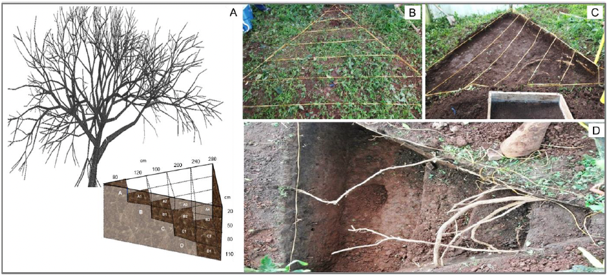
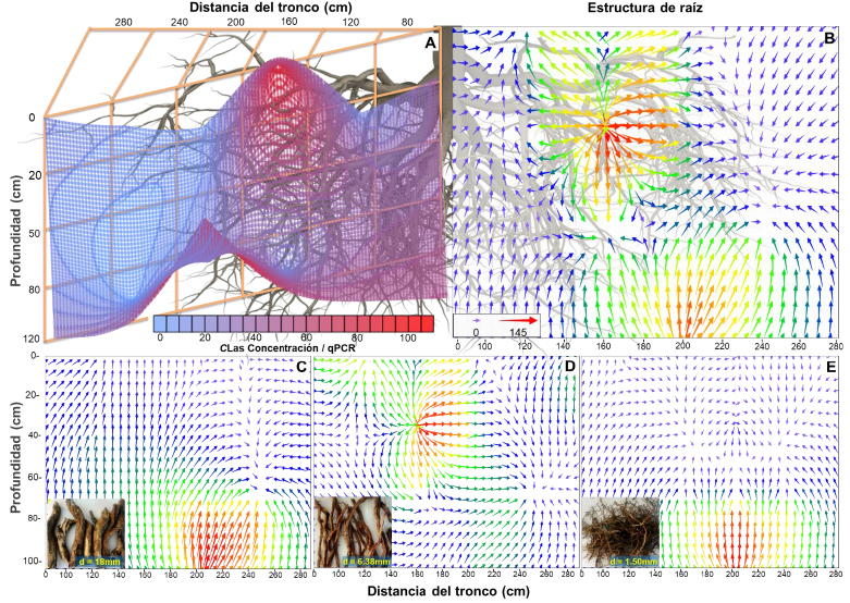
Materials and Methods. Two out of three trees (A1-A3) (>30 years old) of asymptomatic Valencia sweet orange trees positive for CLas, identified through official diagnostic protocols in San José Acateno, Puebla, were confined. On trees A2 and A3, 13 and 16 secondary branches were marked, with a total of 112 and 108 foliage/samples per month, respectively (total 1540 samples), obtained from the distal and proximal parts of each branch. Seven monthly samplings of plant material were carried out between May and December. At the root level, three categories were sampled by diameter/function and by a combination of six distal trunk sectors (0 - 280 cm) by 4 in depth (0 x 110 cm) with a total of 111 samples. DNA extraction was performed with 2% CTAB and quantification by qPCR (oligos, HLBp/HLBas).
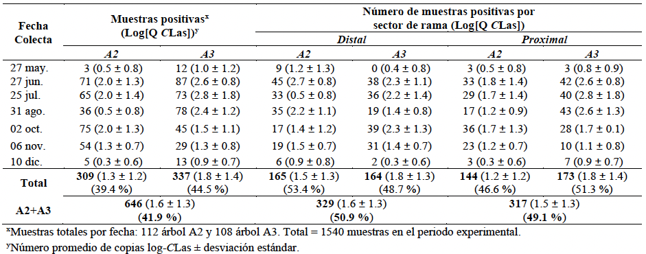
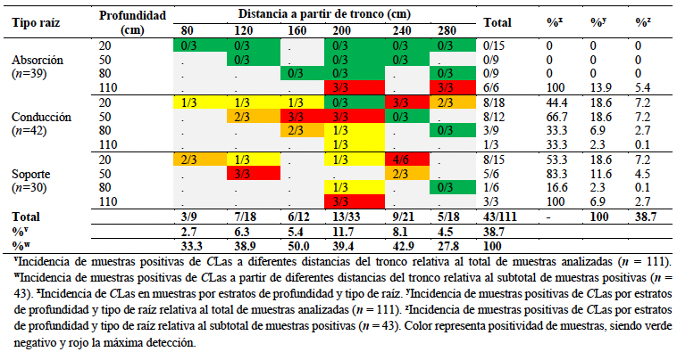
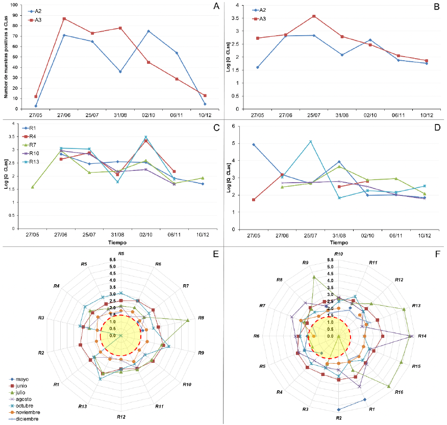
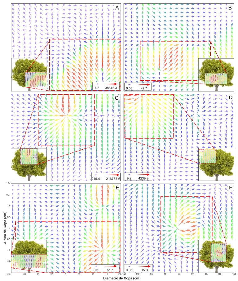
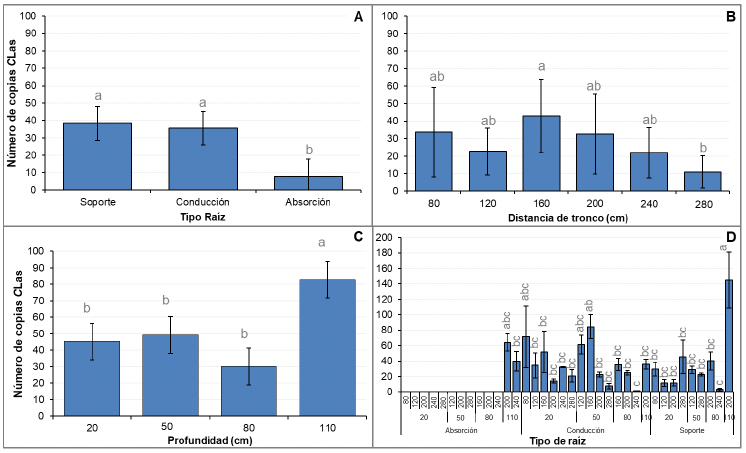
Results. In canopy, the incidence / branche in A3 was 52.2% (Ct = 22.7 – 34.9), and in A2 47.8% (Ct = 23.4 –35), with no statistical difference (p = 0.48). The inoculum load was 1.6 (±1.3) log-CLas, equivalent to 109 copies/reaction in 100 mL (range in number of copies 6.3 to 68,870.7). CLas did not differ between distal (1.6 log) and proximal (1.5 log) samples (p = 0.33). Significative temporal fluctuation was found between June and October in positive samples (37 – 87, p < 0.0001), but not in CLas concentration (1.6 – 3.5 log-CLas, p > 0.72). The distribution of CLas by branch had a temporal effect (p = 0.0001) and by sector (p = 0.07), showing high mobility and seasonality. The maximum concentration (Ymax) was associated with the middle-upper sector of the canopy, and high mobility towards the lower and upper sectors in Yinitial and Yfinal. In roots, 38.7% positivity was found. The detectability of CLas was homogeneous among root types (p < 0.0001) but variable in concentration (p = 0.04). The 47.6, 15.4, and 38.7% incidence was associated with 38.3, 43.4, and 9.5 average CLas copies for support, conduction, and absorption roots, respectively (p = 0.0001). Two sections with higher CLas intensity-concentration (80 – 144 copies) were associated with roots in the middle drip zone (160 – 240 cm) and medium depth strata (50 – 80 cm).
Conclusion. CLas was significantly and consistently detected in plant and root tissue throughout the experimental period, but at fluctuating, non-incremental concentration, analogous between trees A2 and A3. The wide canopy-root distribution suggests a chronic systemic effect. Consequently, the incubation period does not determine the asymptomatic condition in the sweet orange/sour orange productive system investigated. Other factors, e.g. inherent to pathogen variability, could be involved. These results contribute to optimizing a surveillance-monitoring system considering seasonality, zone-branch, sector-canopy and type of tissue, with a greater probability of detection of CLas even in an eventual asymptomatic condition and generate new insights for Clas management. This is the first work that analyzes PI mechanistically, at canopy and root level, in initial detections in Mexico of asymptomatic-HLB in productive adult trees of Valencia C. sinensis / C. aurantium under field conditions.
Share this link via
Or copy link


byNahomy Monserrat Escalera Mares, Juan Daniel Lira Morales, Edith Salazar Villa, Lorena Molina Cárdenas, Indira Rojo Báez*
Received: 10/July/2024 – Published: 27/December/2025
DOI: https://doi.org/10.18781/R.MEX.FIT.2024-32
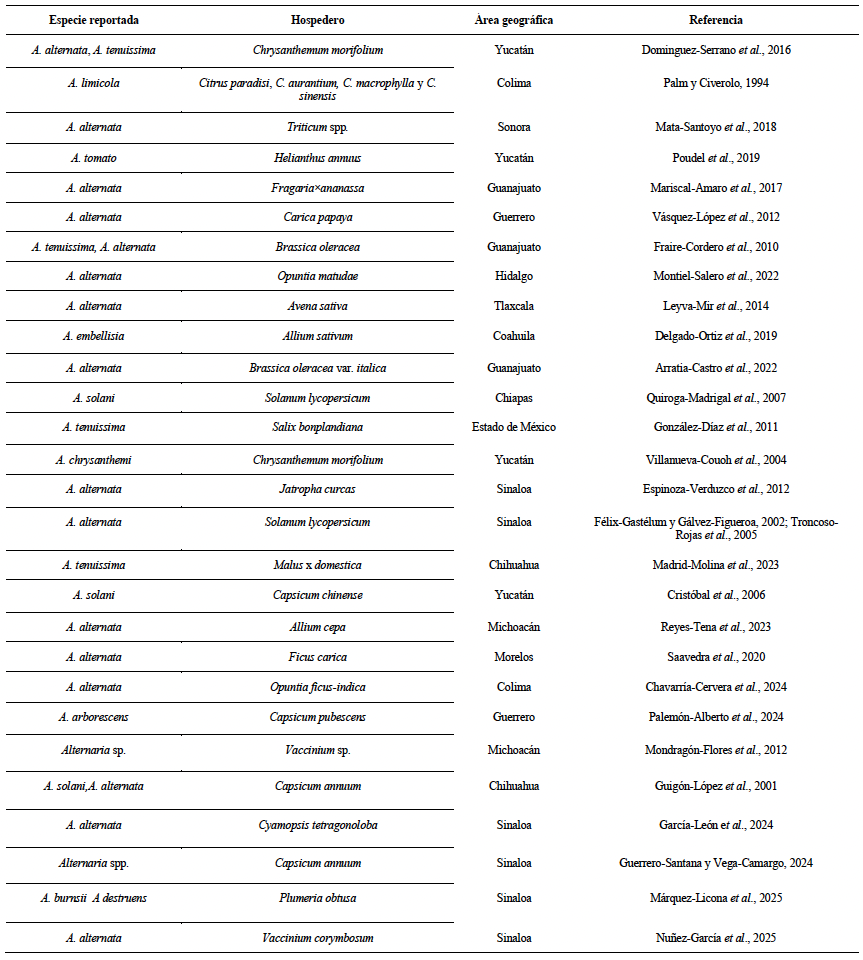
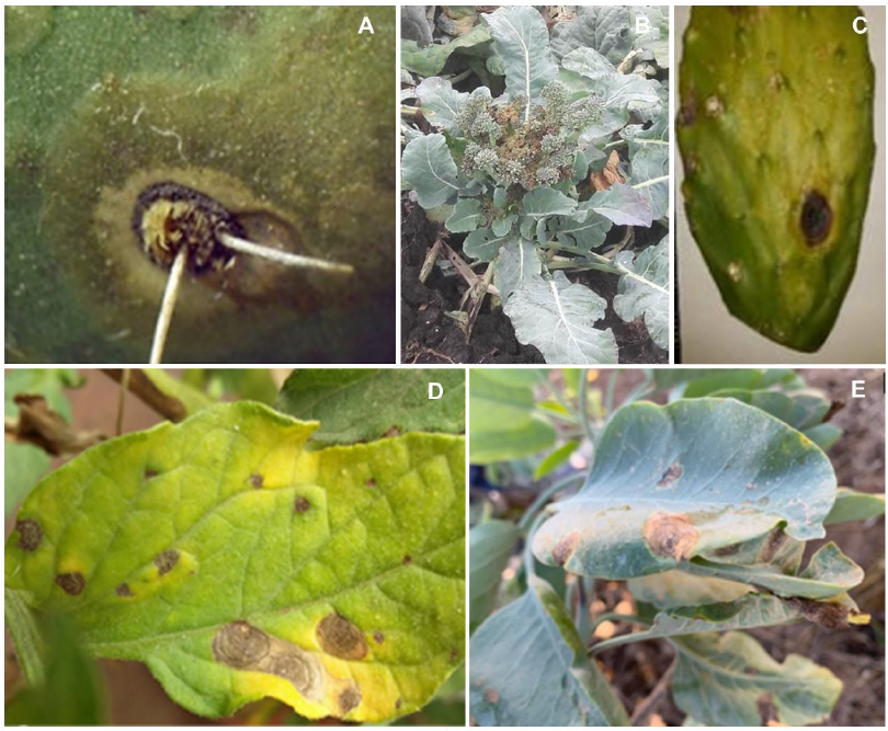
Abstract Justification. Within the genus Alternaria, several phytopathogenic species stand out for causing diseases such as leaf spot and blight in hosts including fruit trees, cereals, vegetables, and ornamental plants, resulting in significant economic losses. Despite this issue, information on the genus Alternaria in Mexico is limited and scattered. Therefore, the objective of this review was to describe the current status of the genus Alternaria in Mexico through a compilation of published studies on the characterization of reported pathogenic species, the diseases they cause in plants, and the various control strategies employed.
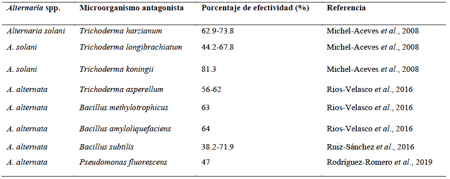
Theoretical and experimental framework. Traditional identification of the genus Alternaria is based on morphological characteristics observed in culture; however, intra- and interspecific variability limits its accuracy. Consequently, molecular tools such as PCR, sequencing, and phylogenetic analysis are used to distinguish species and establish more precise evolutionary relationships. In Mexico, this genus affects more than 20 agricultural and ornamental hosts across 15 states, causing diseases in chrysanthemum, papaya, cacti, broccoli, pepper, and tomato, with incidences exceeding 40% in some crops. Its management requires an integrated approach combining accurate diagnosis, rational use of fungicides, strategies based on crop phenology and inoculum density, and sustainable alternatives such as biological control, plant extracts, microalgae, and nanoparticles, which have shown high efficacy and reduced environmental impact.
Conclusions and Perspectives. In Mexico, nine species have been reported affecting 23 cultivated hosts, although information gaps remain for ornamental, forest, and native species. The main challenges for their control stem from incorrect disease diagnosis, which limits the application of effective strategies.
Share this link via
Or copy link


byMiguel Angel Angulo Escalante, Isabel Cruz Lachica, Edith Salazar Villa, Indira Rojo Báez, Federico Soto Landeros, Isidro Márquez Zequera, Karla Marina Báez Parra*
Received: 16/October/2024 – Published: 22/December/2025
DOI: https://doi.org/10.18781/R.MEX.FIT.2410-3
Abstract Background/Objective. Metabolites of castor bean (Ricinus communis), such as the alkaloid ricinine, have antibacterial properties with the potential to combat phytopathogenic bacteria such as Xanthomonas perforans, Acidovorax citrulli, Clavibacter michiganensis, and Ralstonia solanacearum, making it a natural alternative to conventional chemicals. Therefore, the objective of this study was to evaluate the antimicrobial activity of methanolic extracts from castor bean leaf accessions and quantify ricinine using HPTLC to determine its effectiveness as a bactericidal agent.
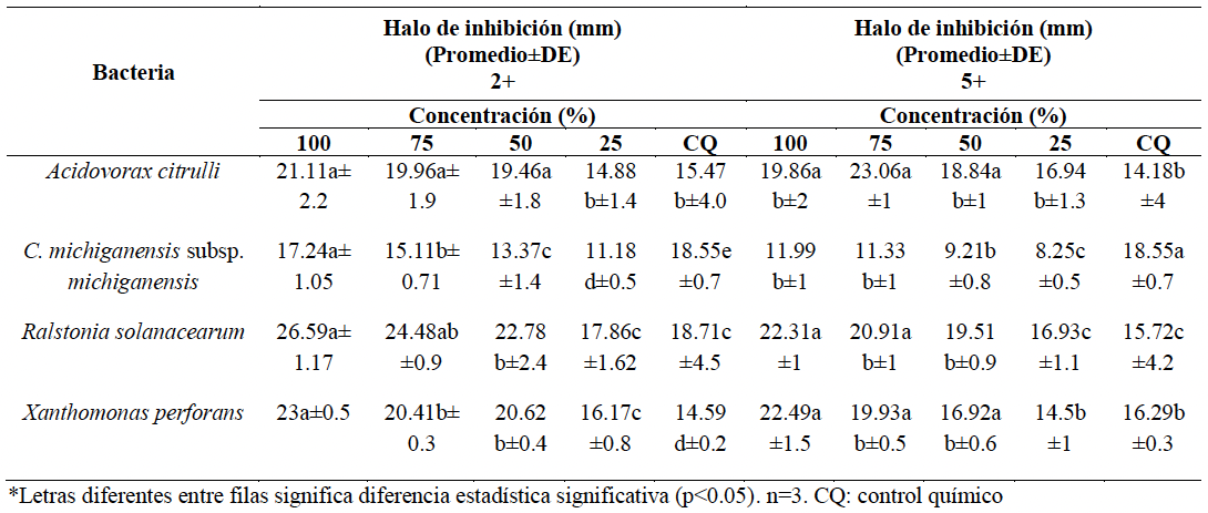
Experimental development. The leaves of nine castor bean accessions were ground and sieved to prepare methanolic extracts coded as: 1+, 2+, 4+, 5+, 6+, 7+, 8+, 9+, and 10+. Ricinine in the extracts was quantified by HPTLC. To determine antibacterial activity, the phytopathogenic bacteria Xanthomonas perforans, Acidovorax citrulli, Clavibacter michiganensis subsp. michiganensis, and Ralstonia solanacearum were reactivated and cultured in selective medium with concentrations of the extracts at 100, 75, 50, and 25% and a chemical control Finalbacter® (Gentamicin Sulfate and Oxytetracycline Hydrochloride). The analysis was performed in triplicate.
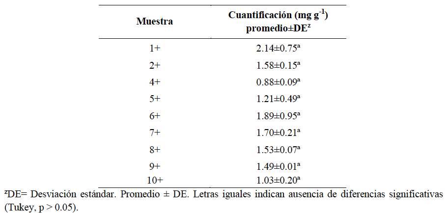
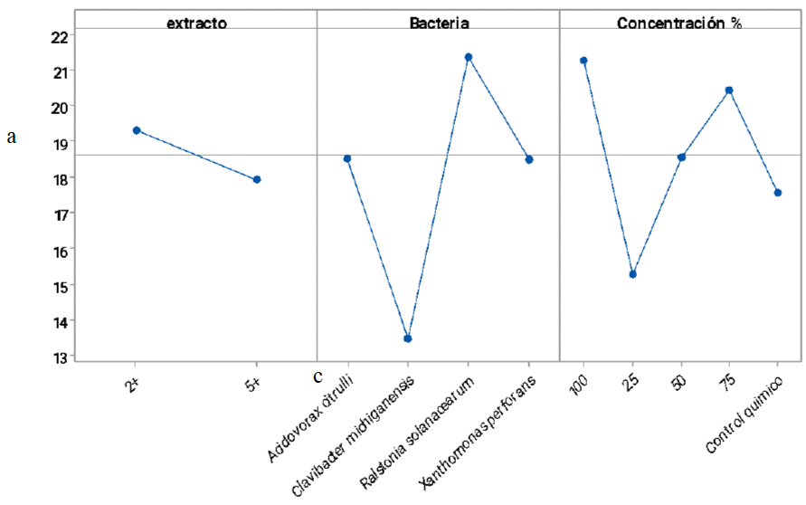
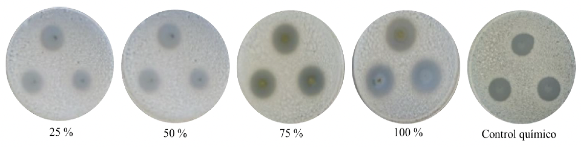
Results. The extracts identified as 2+ and 5+ proved to be the most effective, with inhibition halos of 18.94 mm and 17.23 mm, respectively. Ralstonia solanacearum showed the greatest inhibition (21.6 mm), followed by A. citrulli and X. perforans (19.26 mm each) and C. michiganensis subsp. michiganensis (12.21 mm). The 100% and 75% concentrations showed the largest inhibition halos (20.6 mm and 19.8 mm), compared to the lower concentrations and the chemical control. Ricinine concentrations ranging from 0.88 to 2.14 mg g⁻¹ were quantified.
Conclusion. Accessions 2+ and 5+ of Ricinus communis showed potential as a natural option for controlling phytopathogenic bacteria, with an average inhibition zone of 19.31 mm and 17.92 mm, respectively.
Share this link via
Or copy link
byJosé Francisco Ramírez Dávila, Fidel Lara Vázquez*, Dulce Karen Figueroa Figueroa, María Teresa Lugo Coyote
Received: 14/March/2025 – Published: 20/December/2025
DOI: https://doi.org/10.18781/R.MEX.FIT.2212-5
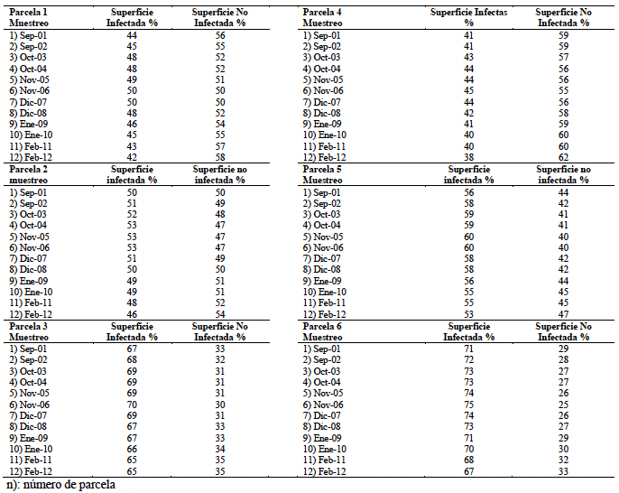
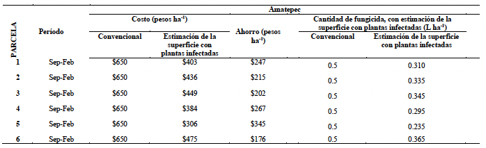
Abstract Background/Objective. Coffee production in the State of Mexico, particularly in the municipality of Amatepec, is threatened by several diseases, including iron spot caused by Mycosphaerella coffeicola. This foliar disease negatively affects coffee yield and quality. The objective of this study was to model the spatial distribution of iron spot in coffee plantations in Amatepec using classical statistics and geostatistics, estimating the infected area to economically evaluate management strategies.
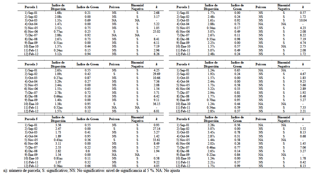
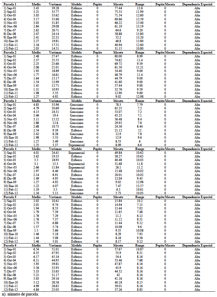
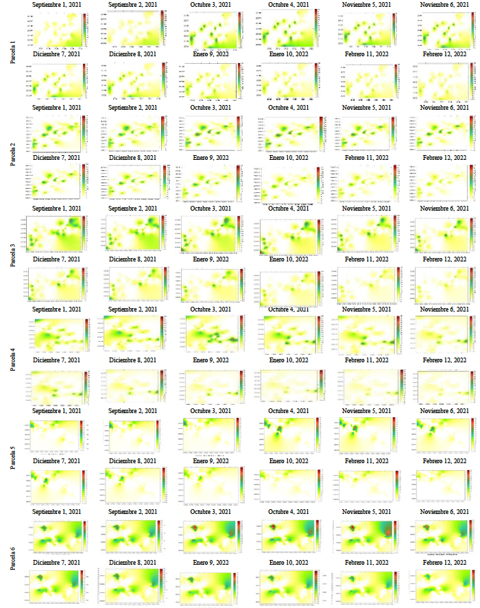
Experimental development. In six coffee plots planted with the cultivars Caturra and Typica, 100 plants per plot were georeferenced using a quadrant sampling method and monitored biweekly for six months. From each plant, 36 leaves were sampled considering the four cardinal points and three canopy strata (upper, middle, and lower). Data were analyzed using classical statistics (dispersion and Green’s indices, Poisson and Negative Binomial distributions) and geostatistics (theoretical semivariograms and Ordinary Kriging).
Results. Classical statistics indicated an aggregated distribution of the disease but lacked sufficient spatial precision. Geostatistical analysis generated theoretical semivariograms (Gaussian, Spherical, and Exponential models) and Ordinary Kriging maps that clearly visualized disease distribution. This approach allowed the identification of infection foci and a reliable estimation of the infected area, supporting precision management strategies.
Conclusion. Geostatistics provided greater precision than classical statistics for spatial analysis of iron spot. Ordinary Kriging maps identified areas with and without disease presence, enabling more efficient and targeted management actions.
Share this link via
Or copy link
byKarla Marina Báez Parra, Edith Salazar Villa, Josefina León Félix, Miguel Ángel Angulo Escalante, Indira Rojo Báez*
Received: 28/July/2024 – Published: 16/December/2025
DOI: https://doi.org/10.18781/R.MEX.FIT.2407-3
Abstract Background/Objective. Biofertilizers exist that contain microorganisms beneficial to plants and the environment. However, some commercial products provide little information about the species they use. Therefore, this study aimed to identify, culturally, morphologically, morphometrically, and molecularly, the Trichoderma species present in the BioHumisol® fertilizer used in agriculture in Culiacán and Navolato, Sinaloa, México.
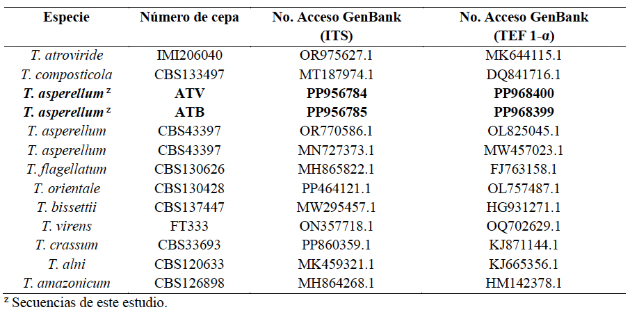
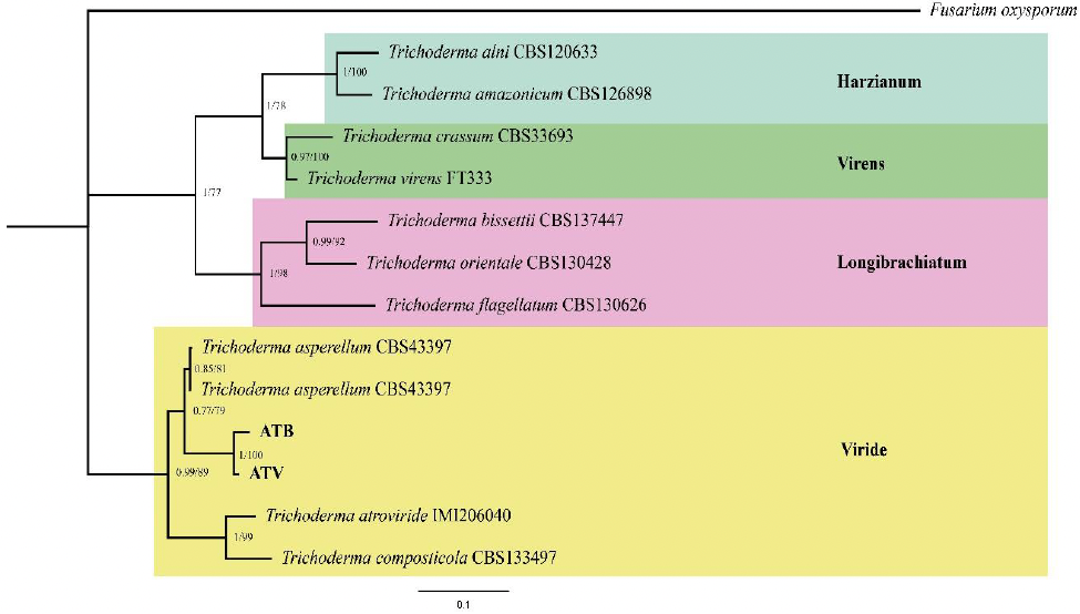
Experimental development. Fungal colonies were isolated and purified from the BioHumisol® liquid biofertilizer. For cultural, morphological, and morphometric identification, three Petri dishes were used, each containing one fungal isolate. The following characteristics were recorded: color, mycelium shape and arrangement in the culture medium, days of sporulation, and the size and shape of conidia, phialides, and chlamydospores. The mycelial growth rate was also recorded. At the molecular level, the ITS region and the TEF-1 α gene were amplified. The sequences were registered in GenBank with the accession numbers: PP956784, PP956785 for ITS; PP968400, PP968399 for TEF-1 α. Phylogenetic analysis of the concatenated sequences was performed using the maximum likelihood and Bayesian inference method with a bootstrap of 1000 replicates.
Results. The fungal colonies presented hyaline, branched, non-whorled conidiophores; phialides either single or in groups with terminal branches worth average measurements of 4.1-6.2 × 9.7-15.3 μm; unicellular ovoid conidia with average dimensions of 3.6-5.09 × 3.27-6.41 μm and subglobose intercalary and terminal chlamydospores measuring 21.41 × 21.72 μm consistent with Trichoderma. Currently accurate identification requires cultural, morphological and morphometric characterization, complemented by molecular characterization. BLAST analysis of partial ITS sequences showed 99% homology, and the TEF -1 α gene showed 100% homology with T. asperellum. Phylogenetic analyses confirm genetic proximity to species of the Viride clade.
Conclusion. Based on the cultural, morphological, morphometric and molecular characterization, the species found in the liquid biofertilizer BioHumisol® is Trichoderma asperellum.
Share this link via
Or copy link